
Urinary System I. Functions of the Urinary System







Description: Urinary System I. Functions of the Urinary System A. Maintain Water Balance B. Maintain Salt Balance C. Maintain pH H D. Excretion of metabolic (biochemical) waste products urea (breakdown product of proteins) and uric acid Urea
Related Topics
Download Presentation
"Urinary System I. Functions of the Urinary System" is the property of its rightful owner. Permission is granted to download and print the materials on this website for personal, non-commercial use only, and to display it on your personal computer provided you do not modify the materials and that you retain all copyright notices contained in the materials. By downloading content from our website, you accept the terms of this agreement.
Presentation Transcript
slide1. Urinary System<br>
slide2. I. Functions of the Urinary System A. Maintain Water Balance
B. Maintain Salt Balance
C. Maintain pH [H+]
D. Excretion of metabolic (biochemical) waste products = urea (breakdown product of proteins) and uric acid<br>
slide3. Urea – major waste product in the body
Uric acid – gouty arthritis or gout
- formed from the breakdown of nucleic acids
feces = food residues<br>
slide4. Regions of the Urinary System A. Kidneys
B. Ureters
C. Urinary Bladder
D. Urethral Canal (urethra)<br>
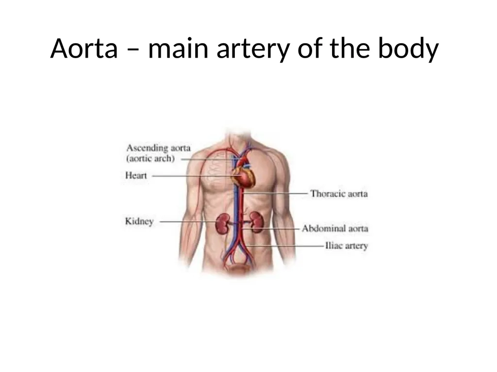
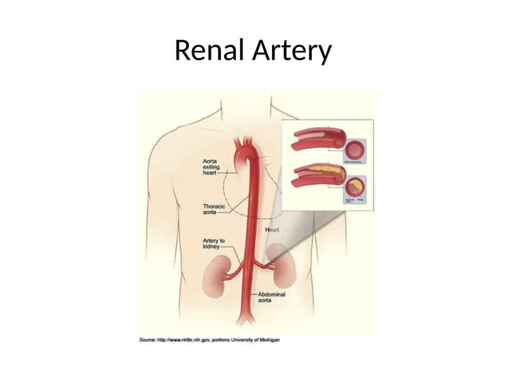
slide5. Aorta – main artery of the body<br>
slide6. Renal Artery<br>
slide7. Purified blood leaves the kidney through the renal vein<br>
slide8. Renal vein = lower in waste products, lower in oxygen
Renal artery = higher oxygen
Most organs in the body are pink because of the number of blood vessels and present in the vessels are red blood cells other organs that are not pink and most other cells/tissues are transparent or has no color<br>
slide9. The Kidneys A. reddish-brown organs located retroperitoneally
(the reddish color is due to the vascularity of the kidneys)
B. the kidneys extend from about T-12 to L-3, with the right kidney located slightly lower than the left kidney<br>
slide10. C. the kidneys are surrounded by renal capsule and perirenal fat – the fat is for protecting the kidney
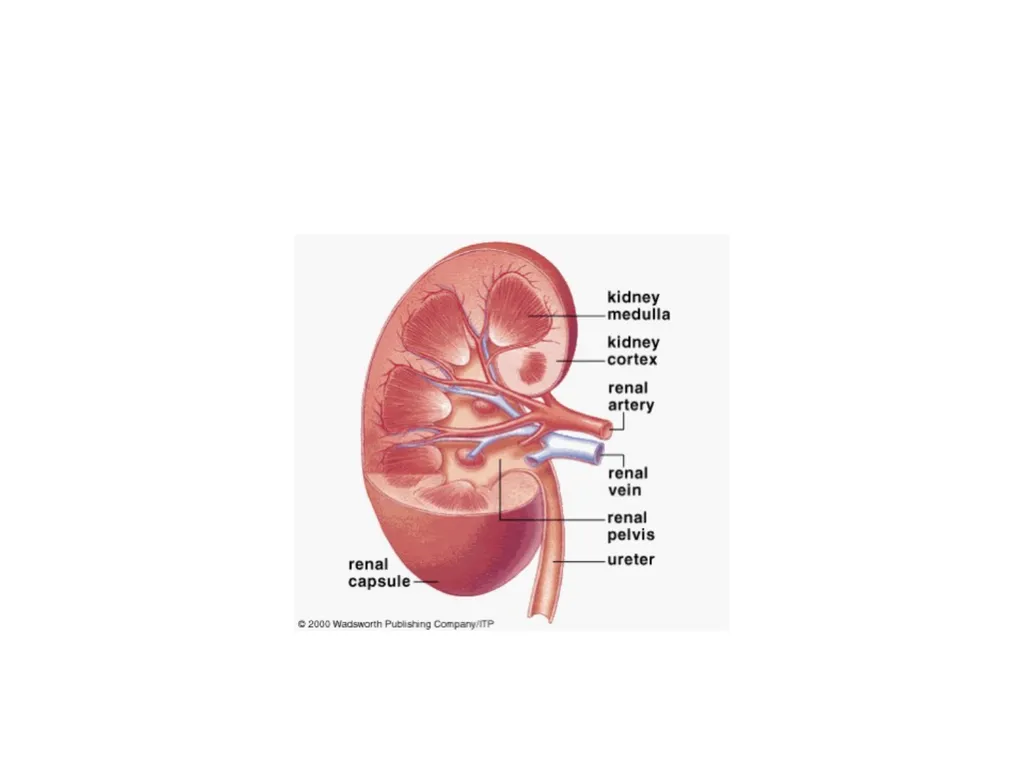
D. renal cortex
“Note: interlobar arteries and veins extend through the renal columns of the cortex dividing the kidneys into lobes<br>
slide11. E. Renal Medulla (Medullary Pyramids)
Note: the urine collecting ducts pass downward through the renal medulla (pyramids) and open into the calyces of the ureter<br>
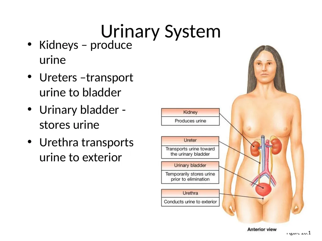
slide17. Figure 26.1 Urinary System Kidneys – produce urine
Ureters –transport urine to bladder
Urinary bladder - stores urine
Urethra transports urine to exterior<br>
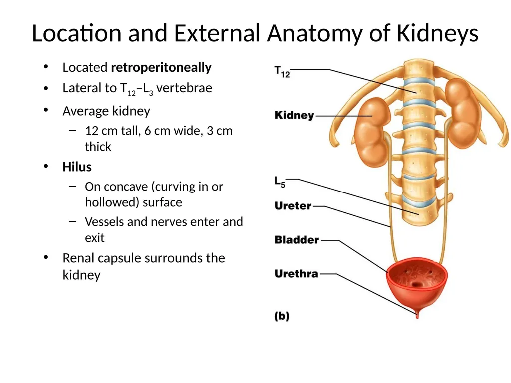
slide18. Location and External Anatomy of Kidneys Located retroperitoneally
Lateral to T12–L3 vertebrae
Average kidney
12 cm tall, 6 cm wide, 3 cm thick
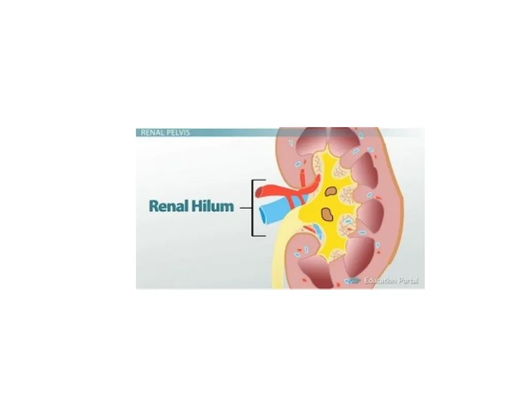
Hilus
On concave (curving in or hollowed) surface
Vessels and nerves enter and exit
Renal capsule surrounds the kidney<br>
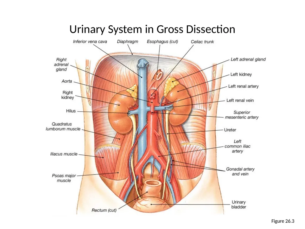
slide19. Figure 26.3 Urinary System in Gross Dissection<br>
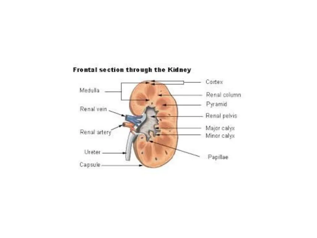
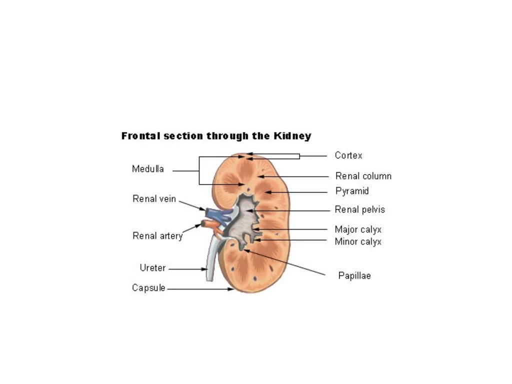
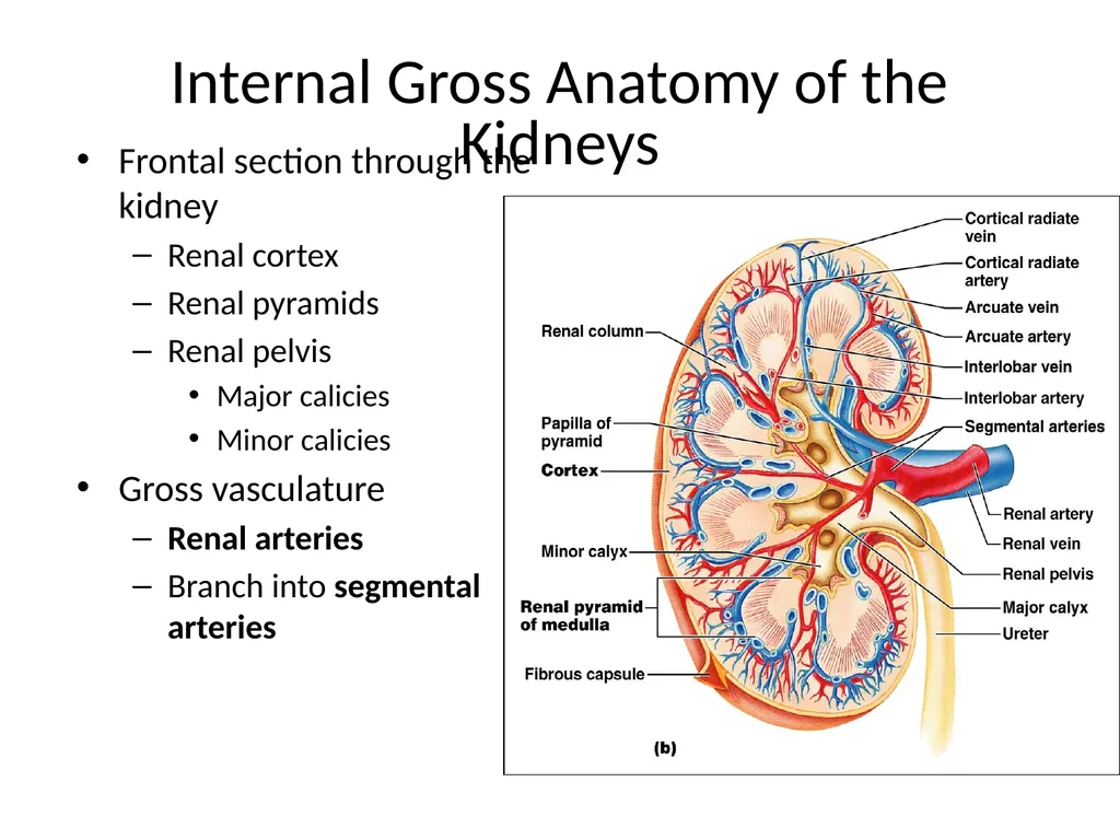
slide20. Internal Gross Anatomy of the Kidneys Frontal section through the kidney
Renal cortex
Renal pyramids
Renal pelvis
Major calicies
Minor calicies
Gross vasculature
Renal arteries
Branch into segmental arteries<br>
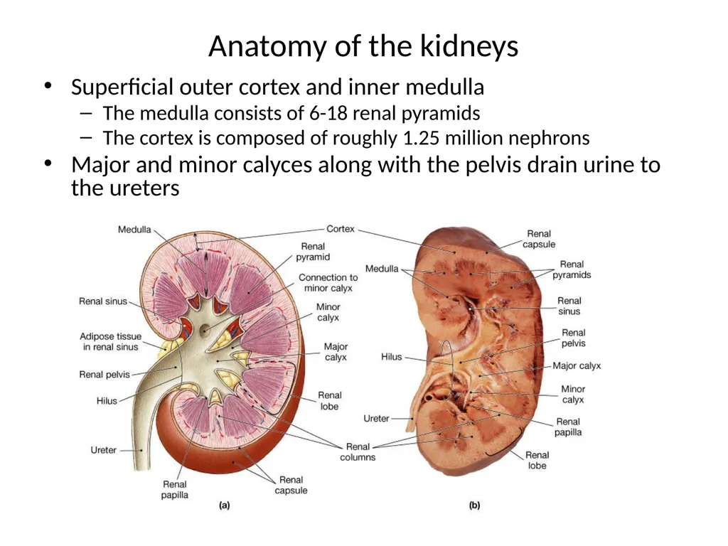
slide21. Anatomy of the kidneys Superficial outer cortex and inner medulla
The medulla consists of 6-18 renal pyramids
The cortex is composed of roughly 1.25 million nephrons
Major and minor calyces along with the pelvis drain urine to the ureters<br>
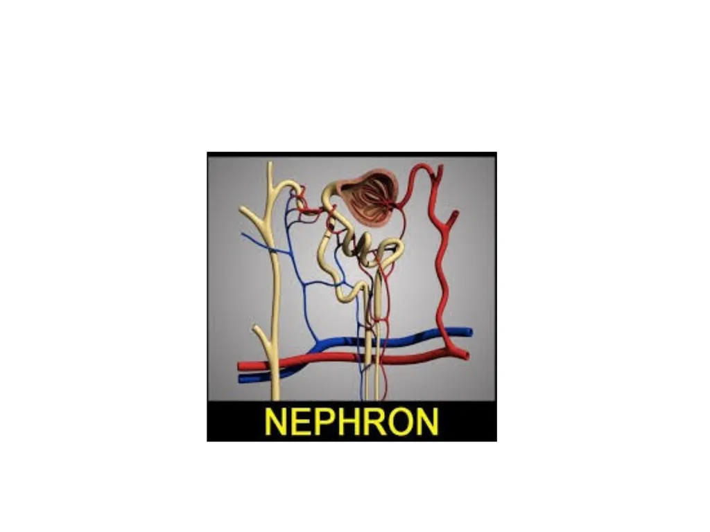
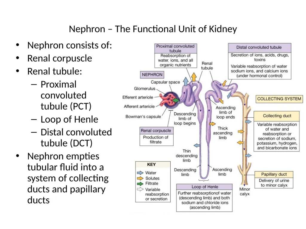
slide22. Nephron – The Functional Unit of Kidney Nephron consists of:
Renal corpuscle
Renal tubule:
Proximal convoluted tubule (PCT)
Loop of Henle
Distal convoluted tubule (DCT)
Nephron empties tubular fluid into a system of collecting ducts and papillary ducts<br>
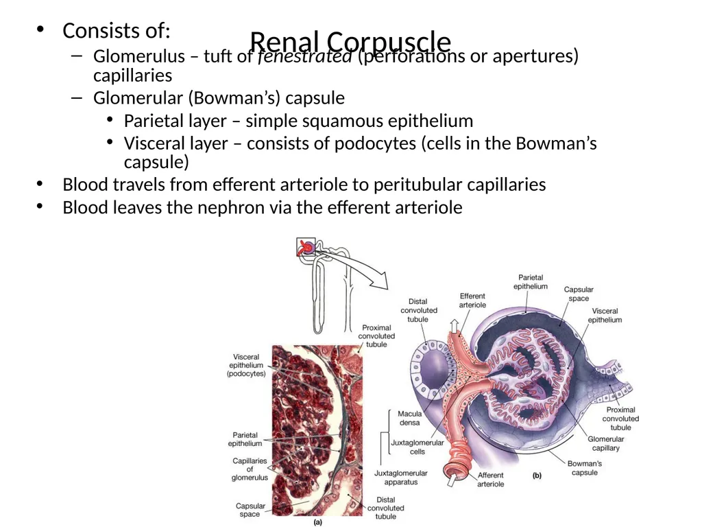
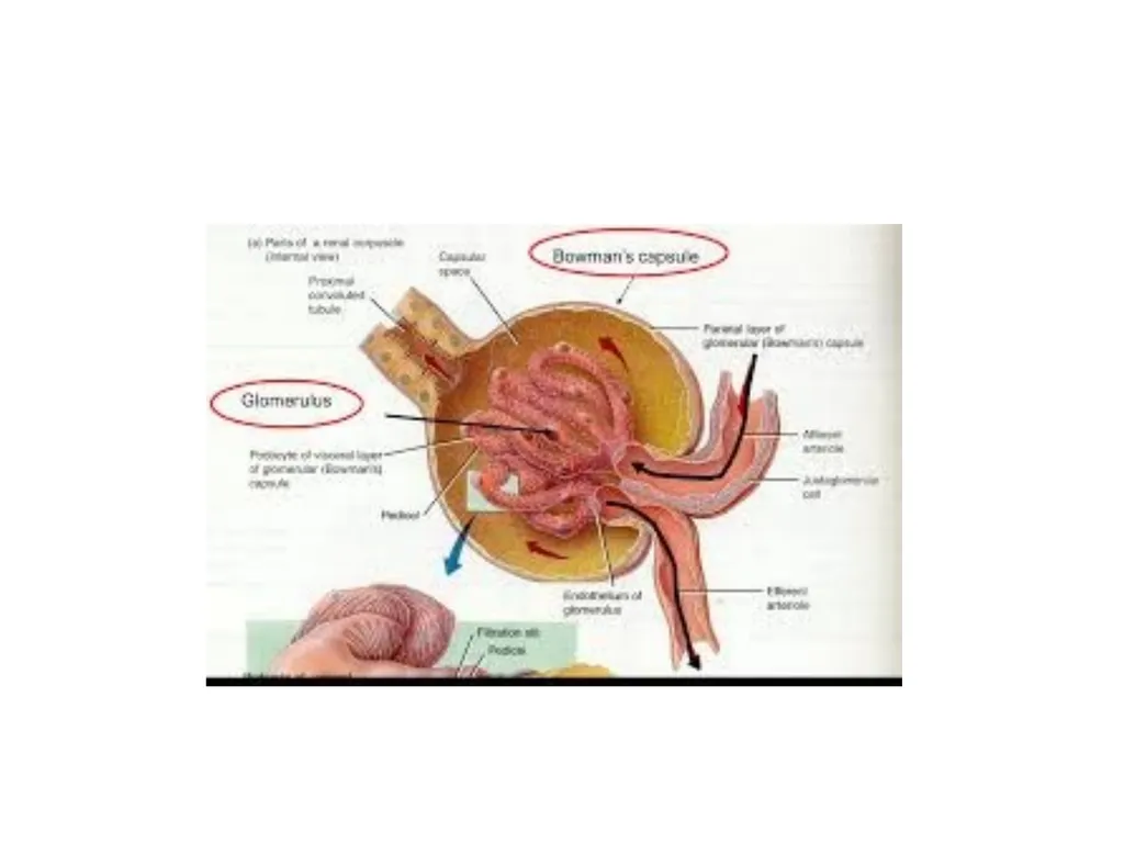
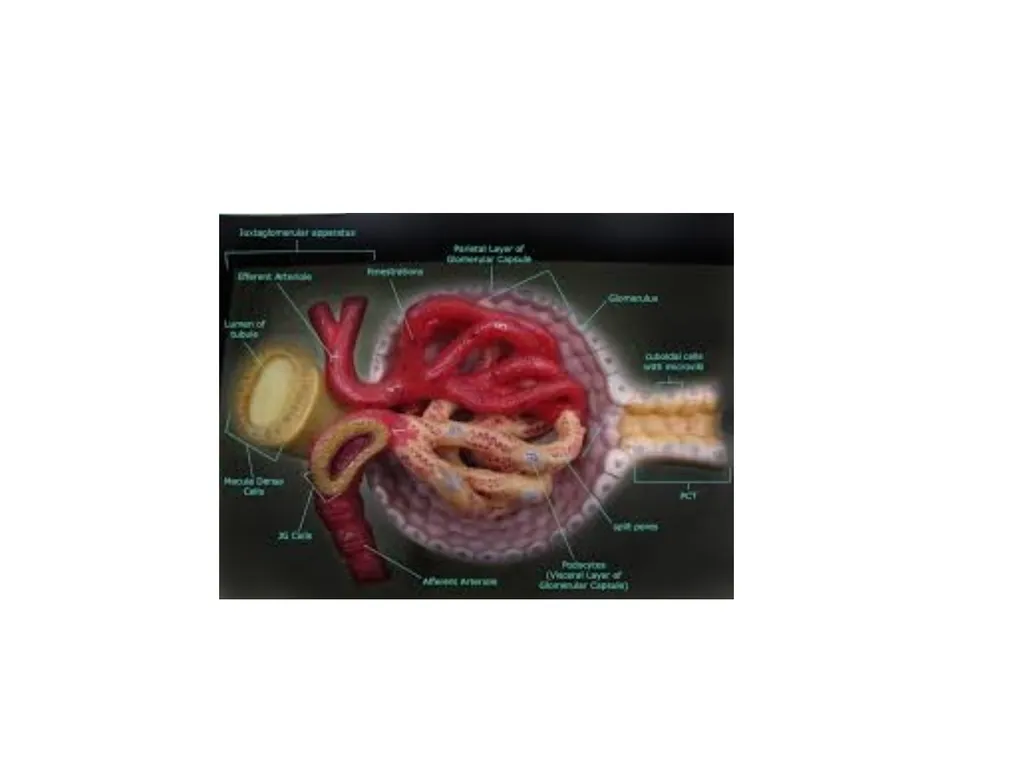
slide23. Renal Corpuscle Consists of:
Glomerulus – tuft of fenestrated (perforations or apertures) capillaries
Glomerular (Bowman’s) capsule
Parietal layer – simple squamous epithelium
Visceral layer – consists of podocytes (cells in the Bowman’s capsule)
Blood travels from efferent arteriole to peritubular capillaries
Blood leaves the nephron via the efferent arteriole<br>
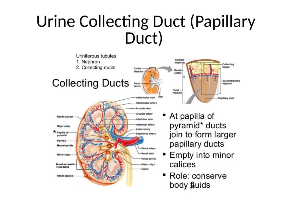
slide24. Urine Collecting Duct (Papillary Duct)<br>
slide25. Blood supply to the kidney Blood flow to the kidney involves a portal system
Renal Artery Interlobar Artery Arcuate Artery Interlobar Artery Afferent Glomerular Arteriole Glomerular Capillaries or Capillary Bed (Glomerulus) Efferent Glomerular Arteriole (Portal Vessel)
Peritubular Capillaries (Capillary Bed) & Vasa Recta Interlobar Vein Arcuate Vein<br>
slide26. Most locations in the body are: Arteries Capillary Bed Vein
But there are 3 places in our body wherein we have what we call portal systems
What are portal systems?
Portal system – 2 capillary beds in between
Artery Capillary Bed Portal Vessel
Capillary Bed Vein<br>
slide27. Where can you find portal systems? 1. Hepatic Portal System
2. Kidney
3. Hypothalamic-Pituitary System
Example in the liver:
Arteries Capillaries Hepatic Portal Vein
Liver
Or Digestive Tract Arteries Capillaries
Hepatic Portal Vein Capillaries Vein<br>
slide28. Glomerular Capsule or Bowman’s Capsule – named after Dr. Bowman<br>
slide29. Microanatomy of the Kidney A. The Nephrons (Renal Tubules) – cuboidal epithelial cells with microvilli
1. Glomerular (Bowman’s) Capsule
a. encloses the glomerular capillaries
1. glomerular filtration of blood – some of the fluid in the bloodstream filters out into the glomerular capsule<br>
slide30. 2. Proximal Convoluted Tubule – location for tubular reabsorption
- the “good stuff” (sugars & amino acids) is reabsorbed back into the bloodstream (going from the proximal convoluted tubule into the peritubular capillaries)<br>
slide33. Loop of Henle (“Nephron Loop”)
- located in the renal medulla
- consists of a descending portion and an ascending portion (limb)
Distal Convoluted Tubule – location for tubular secretion – certain specific chemicals are transported out of the peritubular capillaries into the distal convoluted tubule like excess H+, excess NaCl, pencillin antibiotics<br>
slide34. The Urine Collecting Ducts (Papillary Ducts) – the nephrons empty into the urine collecting ducts, which conduct the urine down to the renal calyces of the ureter
The Ureters – are a 10-inch long muscular tube that conduct urine from the kidneys to the urinary bladder by peristalsis<br>
slide35. “Study without thought is labor lost; thought without study is perilous. By nature men are nearly alike, but through experience they grow wide apart. Those who are born wise are the highest type of men; those who become wise through learning come next; those who are dull-witted and yet strive to learn come after that. Those who are dull-witted and yet make no effort to learn are the lowest type of men” – Confucius<br>
slide2. I. Functions of the Urinary System A. Maintain Water Balance
B. Maintain Salt Balance
C. Maintain pH [H+]
D. Excretion of metabolic (biochemical) waste products = urea (breakdown product of proteins) and uric acid<br>
slide3. Urea – major waste product in the body
Uric acid – gouty arthritis or gout
- formed from the breakdown of nucleic acids
feces = food residues<br>
slide4. Regions of the Urinary System A. Kidneys
B. Ureters
C. Urinary Bladder
D. Urethral Canal (urethra)<br>
slide5. Aorta – main artery of the body<br>
slide6. Renal Artery<br>
slide7. Purified blood leaves the kidney through the renal vein<br>
slide8. Renal vein = lower in waste products, lower in oxygen
Renal artery = higher oxygen
Most organs in the body are pink because of the number of blood vessels and present in the vessels are red blood cells other organs that are not pink and most other cells/tissues are transparent or has no color<br>
slide9. The Kidneys A. reddish-brown organs located retroperitoneally
(the reddish color is due to the vascularity of the kidneys)
B. the kidneys extend from about T-12 to L-3, with the right kidney located slightly lower than the left kidney<br>
slide10. C. the kidneys are surrounded by renal capsule and perirenal fat – the fat is for protecting the kidney
D. renal cortex
“Note: interlobar arteries and veins extend through the renal columns of the cortex dividing the kidneys into lobes<br>
slide11. E. Renal Medulla (Medullary Pyramids)
Note: the urine collecting ducts pass downward through the renal medulla (pyramids) and open into the calyces of the ureter<br>
slide17. Figure 26.1 Urinary System Kidneys – produce urine
Ureters –transport urine to bladder
Urinary bladder - stores urine
Urethra transports urine to exterior<br>
slide18. Location and External Anatomy of Kidneys Located retroperitoneally
Lateral to T12–L3 vertebrae
Average kidney
12 cm tall, 6 cm wide, 3 cm thick
Hilus
On concave (curving in or hollowed) surface
Vessels and nerves enter and exit
Renal capsule surrounds the kidney<br>
slide19. Figure 26.3 Urinary System in Gross Dissection<br>
slide20. Internal Gross Anatomy of the Kidneys Frontal section through the kidney
Renal cortex
Renal pyramids
Renal pelvis
Major calicies
Minor calicies
Gross vasculature
Renal arteries
Branch into segmental arteries<br>
slide21. Anatomy of the kidneys Superficial outer cortex and inner medulla
The medulla consists of 6-18 renal pyramids
The cortex is composed of roughly 1.25 million nephrons
Major and minor calyces along with the pelvis drain urine to the ureters<br>
slide22. Nephron – The Functional Unit of Kidney Nephron consists of:
Renal corpuscle
Renal tubule:
Proximal convoluted tubule (PCT)
Loop of Henle
Distal convoluted tubule (DCT)
Nephron empties tubular fluid into a system of collecting ducts and papillary ducts<br>
slide23. Renal Corpuscle Consists of:
Glomerulus – tuft of fenestrated (perforations or apertures) capillaries
Glomerular (Bowman’s) capsule
Parietal layer – simple squamous epithelium
Visceral layer – consists of podocytes (cells in the Bowman’s capsule)
Blood travels from efferent arteriole to peritubular capillaries
Blood leaves the nephron via the efferent arteriole<br>
slide24. Urine Collecting Duct (Papillary Duct)<br>
slide25. Blood supply to the kidney Blood flow to the kidney involves a portal system
Renal Artery Interlobar Artery Arcuate Artery Interlobar Artery Afferent Glomerular Arteriole Glomerular Capillaries or Capillary Bed (Glomerulus) Efferent Glomerular Arteriole (Portal Vessel)
Peritubular Capillaries (Capillary Bed) & Vasa Recta Interlobar Vein Arcuate Vein<br>
slide26. Most locations in the body are: Arteries Capillary Bed Vein
But there are 3 places in our body wherein we have what we call portal systems
What are portal systems?
Portal system – 2 capillary beds in between
Artery Capillary Bed Portal Vessel
Capillary Bed Vein<br>
slide27. Where can you find portal systems? 1. Hepatic Portal System
2. Kidney
3. Hypothalamic-Pituitary System
Example in the liver:
Arteries Capillaries Hepatic Portal Vein
Liver
Or Digestive Tract Arteries Capillaries
Hepatic Portal Vein Capillaries Vein<br>
slide28. Glomerular Capsule or Bowman’s Capsule – named after Dr. Bowman<br>
slide29. Microanatomy of the Kidney A. The Nephrons (Renal Tubules) – cuboidal epithelial cells with microvilli
1. Glomerular (Bowman’s) Capsule
a. encloses the glomerular capillaries
1. glomerular filtration of blood – some of the fluid in the bloodstream filters out into the glomerular capsule<br>
slide30. 2. Proximal Convoluted Tubule – location for tubular reabsorption
- the “good stuff” (sugars & amino acids) is reabsorbed back into the bloodstream (going from the proximal convoluted tubule into the peritubular capillaries)<br>
slide33. Loop of Henle (“Nephron Loop”)
- located in the renal medulla
- consists of a descending portion and an ascending portion (limb)
Distal Convoluted Tubule – location for tubular secretion – certain specific chemicals are transported out of the peritubular capillaries into the distal convoluted tubule like excess H+, excess NaCl, pencillin antibiotics<br>
slide34. The Urine Collecting Ducts (Papillary Ducts) – the nephrons empty into the urine collecting ducts, which conduct the urine down to the renal calyces of the ureter
The Ureters – are a 10-inch long muscular tube that conduct urine from the kidneys to the urinary bladder by peristalsis<br>
slide35. “Study without thought is labor lost; thought without study is perilous. By nature men are nearly alike, but through experience they grow wide apart. Those who are born wise are the highest type of men; those who become wise through learning come next; those who are dull-witted and yet strive to learn come after that. Those who are dull-witted and yet make no effort to learn are the lowest type of men” – Confucius<br>