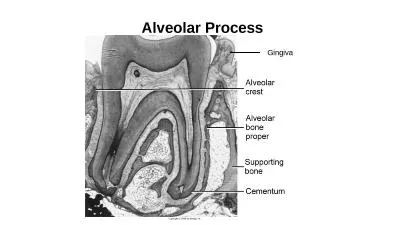
/
Alveolar bone: Is that part of upper/lower jaw that responsible for carrying the teeth Alveolar bone: Is that part of upper/lower jaw that responsible for carrying the teeth

Alveolar bone: Is that part of upper/lower jaw that responsible for carrying the teeth - PowerPoint Presentation

violet
violet . @violet
Follow
344 views
Uploaded On 2022-06-07

Alveolar bone: Is that part of upper/lower jaw that responsible for carrying the teeth - PPT Presentation

1 Houses the roots of teethAnchors the roots of teeth to the alveoli which is achieved by the insertion of Sharpeys fibers into the alveolar bone proper 2Helps to move the teeth for better occlusion ID: 914166

alveolar bone tooth teeth bone alveolar teeth tooth resorption bundle process periodontal proper spongy cortical loss trabeculae plates mesial

Share:

Link:

Embed:

Download Presentation from below link

Download Presentation The PPT/PDF document "Alveolar bone: Is that part of upper/low..." is the property of its rightful owner. Permission is granted to download and print the materials on this web site for personal, non-commercial use only, and to display it on your personal computer provided you do not modify the materials and that you retain all copyright notices contained in the materials. By downloading content from our website, you accept the terms of this agreement.


Presentation Transcript

Slide1

Alveolar bone:

Slide2

Slide3

Is that part of upper/lower jaw that responsible for carrying the teeth and support them. In addition to provide the attachment of some muscles of the face and mastication.

Slide4

Slide5

1- Houses the roots of teeth:Anchors

the roots of teeth to the alveoli, which is achieved by the insertion of

Sharpey’s

fibers into the alveolar bone proper

2-Helps to move the teeth for better occlusion.

3-Helps to absorb and distribute occlusal forces generated during tooth contact.

4-Supplies vessels to periodontal ligament.

5- Houses and protects developing permanent teeth, while supporting primary teeth.

6-Organizes eruption of primary and permanent teeth.

Functions of alveolar bone are:

Slide6

Slide7

Near the end of the second month of fetal life, the maxilla as well as the mandible form a groove that is open towards the surface of the oral cavity.

Tooth germs develop within the bony structures at late bell stage. Bony septa and bony bridge begin to form and separate the individual tooth germs from one another, keeping individual tooth germs in clearly outlined bony compartments.

DEVELOPMENT OF ALVEOLAR PROCESS

Slide8

Tooth germ movement occurs by bone remodeling of bony compartment through bone resorption and bone deposition.

The major changes in the alveolar process begin to occur with development of roots and tooth eruption. As roots develop, the alveolar process increases in height. Also, the cells in the dental follicle start to differentiate into periodontal ligament and cementum.

Slide9

At the same time, some cells in the dental follicle differentiate into

osteoblasts

and form alveolar bone proper.

Hence, an alveolar process develops only during the eruption of the teeth. It is important to realize that, during growth, part of the alveolar process is gradually incorporated into the maxillary or mandibular body while it grows at a fairly rapid rate at its free borders.

The alveolar process gradually diminishes in height after the loss of teeth.

Slide10

Two parts of the alveolar process can be distinguished, the alveolar bone proper and the supporting alveolar bone.

a-Alveolar bone proper

The alveolar bone proper consists partly of lamellated and partly of bundle bone. It surrounds the root of the tooth and gives attachment to principal fibers of the periodontal ligament.

STRUCTURE OF THE ALVEOLAR BONE

Slide11

The lamellar bone contains osteons

each of which has a blood vessel in a

haversian

canal. Blood vessel is surrounded by concentric lamellae to form

osteon

.

Some lamellae of the lamellated bone are arranged roughly parallel to the surface of the adjacent marrow spaces, whereas others form

haversian

systems.

1-Lamellated bone

Slide12

Slide13

Bundle bone is that bone in which the principal fibers of the periodontal ligament are anchored. The term ‘bundle’ was chosen, because, the bundles of the principal fibers continue into the bone as

Sharpey’s

fibers. The bundle bone is characterized by the scarcity of the fibrils in the intercellular substance. These fibrils, more over, are all arranged at right angles to

Sharpey’s

fibers. Bundle bone is formed in areas of recent bone apposition.

2-Bundle bone:

Slide14

Radiographically, it is also referred to as the lamina dura, because, of increased

radiopacity

, which is due to the presence of thick bone without

trabeculations

, that X-rays must penetrate and not to any increased mineral content.

The alveolar bone proper, which forms the inner wall of the socket is perforated by many openings that carry branches of the

interalveolar

nerves and blood vessels into the periodontal ligament, and it is therefore called the

cribriform plate

.

Slide15

Bone between the teeth is called

interdental

septum

and is composed entirely of cribriform plate. The

interdental

and

interradicular

septa contain the perforating canals of

Zuckerkandl

and Hirschfeld (nutrient canals) which house the interdental and

interradicular

arteries, veins, lymph vessels and nerves.

Slide16

(a) Cortical plates (b) Spongy bone

a-Cortical plates

Cortical plates consist of compact bone and form the outer and inner plates of the alveolar processes. The cortical plates, continuous with the compact layers of the maxillary and mandibular body, are generally much thinner in the maxilla, than in the mandible.

They are thickest in the premolar and molar region of the lower jaw, especially on the buccal side. In the maxilla, the outer cortical plate is perforated by many small openings through which blood and lymph vessels pass. In the region of the anterior teeth of both jaws, the supporting bone usually is very thin.

The supporting alveolar bone consists of two parts:

Slide17

No spongy bone is found here, and the cortical plate is fused with the alveolar bone proper. Both cribriform plate and cortical plate are compact bone separated by spongy bone.

Histologically, the cortical plates consist of longitudinal lamellae and

haversian

systems .In the lower jaw, circumferential or basic lamellae reach from the body of the mandible into the cortical plates.

Slide18

b-Spongy bone

Spongy bone fills the area between the cortical plates and the alveolar bone proper. It contains

trabeculae

of lamellar bone. These are surrounded by marrow that is rich in

adipocytes

and

pluripotent

mesenchymal cells. The

trabeculae

contain osteocytes in the interior and

osteoblasts

or osteoclasts on the surface. These

trabeculae

of the spongy bone buttress the functional forces to which alveolar bone proper is exposed. The

cancellous

component in maxilla is more than in the mandible.

Slide19

Type I the interdental

and

interradicular

trabeculae

are regular and horizontal in a ladder like arrangement. The architecture of type I is seen most often in the mandible and fits well into the general idea of a trajectory pattern of spongy bone.

Type II shows irregularly arranged, numerous, delicate

interdental

and

interradicular trabeculae. Type II, although evidently functionally satisfactory, lacks a distinct trajectory pattern, which seems to be compensated for

The of the

spongiosa

of the alveolar process into two main types:

Slide20

by the greater number of trabeculae

in any given area. This arrangement is more common in the maxilla.

Both types show a variation in thickness of

trabeculae

and size of marrow spaces.

Slide21

Mesial drift and continuous tooth eruption elicit remodeling of alveolar bone proper. During the mesial drift of a tooth, bone is apposed on the distal and resorbed on the mesial alveolar wall .The distal wall is made up almost entirely of bundle bone. However, the osteoclasts in the adjacent marrow spaces remove part of the bundle bone, when it reaches a certain thickness.

INTERNAL RECONSTRUCTION OF ALVEOLAR BONE

Slide22

In its place, lamellated bone is deposited. On the mesial alveolar wall of a drifting tooth, the sign of active resorption is the presence of

Howship’s

lacunae

containing

osteoclasts. Bundle bone, however, on this side is always present in some areas but forms merely a thin layer. This is because the mesial drift of a tooth does not occur simply as a bodily movement. Thus resorption does not involve the entire mesial surface of the alveolus at one and the same time

Slide23

Moreover, periods of resorption alternate with periods of rest and repair. It is during these periods of repair that bundle bone is formed, and detached periodontal fibers are again secured. Islands of bundle bone are separated from the lamellated bone by reversal lines that turn their convexities towards the lamellated bone.

Slide24

During these changes, compact bone may be replaced by spongy bone or spongy bone may change into compact bone.

This type of internal reconstruction of bone can be observed in physiologic mesial drift or in orthodontic mesial or distal movement of teeth. In these movements an

interdental

septum shows apposition on one surface and resorption on the other.

The result is a reconstructive shift of the

interdental

septum. Alterations in the structure of the alveolar bone are of great importance in connection with the physiologic eruptive movements of the teeth.

Slide25

These movements are directed

mesioocclusally

. At the alveolar

fundus

the continual apposition of bone can be recognized by resting lines separating parallel layers of bundle bone. When the bundle bone has reached a certain thickness, it is resorbed partly from the marrow spaces and then replaced by lamellated bone or spongy

trabeculae

.

Slide26

In older individuals: – Alveolar sockets appear jagged and uneven.

– The marrow spaces have fatty infiltration.

– The alveolar process in edentulous jaws decreases in size.

– Loss of maxillary bone is accompanied by increase in size of the maxillary sinus.

Internal

trabecular

arrangement is more open, which indicates bone loss.

– The distance between the crest of the alveolar bone and CEJ increases with age—approximately by 2.81 mm.

AGE CHANGES:

Slide27

Bone, although one of the hardest tissues of the human body, is biologically a highly plastic tissue.

Where bone is covered by a

vascularized

connective tissue, it is exceedingly sensitive to pressure, whereas tension acts generally as a stimulus to the production of new bone.

It is this biologic plasticity that enables the orthodontist to move teeth without disrupting their relations to the alveolar bone.

Bone is resorbed on the side of pressure and apposed on the side of tension; thus the entire alveolus is allowed to shift with the tooth.

CLINICAL CONSIDERATIONS

Slide28

At sites of alveolar bone compression, osteoclasts proliferate and initial resorption of the superficial bone takes place.

It is believed that, the initial response may involve

osteoblasts

which can produce

collagenolytic

enzymes to remove a portion of unmineralized extracellular matrix, thereby, facilitating access of

osteoclast

precursors to the bone surface.

Slide29

Osteoblastic cells also produce cytokines and chemokines

, which can attract

monocyte

precursors and promote

osteoclast

differentiation.retraction

or apoptotic death of bone lining cells will expose the mineralized bone surface to osteoclasts. At sites of tension,

osteoblasts

are activated to produce osteoid that subsequently mineralizes to form new bone.

Slide30

The adaptation of bone to function is quantitative as well as qualitative. Whereas, increase in functional forces leads to formation of new bone, decreased function leads to a decrease in the volume of bone. This can be observed in the supporting bone of teeth that have lost their antagonists. Here the spongy bone around the alveolus shows pronounced rarefaction

Slide31

The bone trabeculae are less numerous and very thin

.

The

alveolar bone proper, however, is generally well preserved because it continues to receive some stimuli from the tension of the periodontal tissues. During healing of fractures or extraction wounds, an embryonic type of bone is formed, which only later is replaced by mature bone.

Slide32

The embryonic bone also called immature or coarse

fibrillar

bone

, is characterized, among other aspects, by the greater number, size and irregular arrangement of the

osteocytes

than are found in mature bone

.

The

greater number of cells and the reduced volume of calcified intercellular substance render this immature bone more radiolucent than mature bone.

Slide33

This explains why bony callus cannot be seen in radiographs at a time when

histologic

examination of a fracture reveals a well-developed union between the fragments and why a socket after an extraction wound appears to be empty at a time, when it is almost filled with immature bone. The visibility in radiographs lags 2 or 3 weeks behind actual formation of new bone.

Slide34

The most frequent and harmful change in the alveolar process is that which is associated with periodontal disease. The bone resorption is almost universal, occurs more frequently in posterior teeth, is usually symmetrical, occurs in episodic spurts, is both of the horizontal and vertical type (i.e., occurs from the gingival and tooth side, respectively), and is intimately related to bacterial plaque and pocket formation. It has been shown, for example,

endotoxins

produced by the gram- negative bacteria of the plaque lead to an increase in

cAMP

, which increases the

osteoclastic

activity.

Slide35

Resorption after tooth loss has been shown to follow a predictable pattern. The labial aspect of the alveolar crest is the principal site of resorption, which reduces first in width and later in height. The pattern of resorption is different in the maxilla and mandible. The residual alveolar ridge

resorbs

downward and outward in the mandible, whereas, in the maxilla the resorption is upwards and inwards.

Nontraumatic

loss of anterior maxillary teeth is followed by a progressive loss of bone mainly from the labial side.

Slide36

In the deciduous dentition, loss of a retained second deciduous molar, which has no succedaneous

permanent tooth to replace it, is also associated with bone loss. The cause for resorption of alveolar bone after tooth loss has been assumed to be due to disuse atrophy, decreased blood supply, localized inflammation or unfavorable prosthesis pressure.

Slide37

Alveolar ridge defects and deformities can also be the result of congenital defects, trauma, periodontal disease or surgical ablation, as in the case of tumor surgery. Lamina dura is an important diagnostic landmark in deter- mining health of the periapical tissues. Loss of density usually means infections, inflammation and resorption of bone socket.

Slide38

Traditional treatment methods for promoting bone healing primarily utilize bone grafts or synthetic materials to fill the defects and provide structural support.

Bone grafting to stimulate bone deposition has been used in periodontal surgery.

It involves a surgical procedure to place bone or bone substitute material into a bone defect with the objective of producing new bone and possibly the regeneration of periodontal ligament and cementum.

THERAPEUTIC CONSIDERATIONS

Slide39

1-Autografts utilize the patient’s bone, which can be obtained from intraoral or extraoral

sites. They are the best materials for bone grafting, are very well accepted by the body and may produce the fastest rate of bone growth .

With

autografts

, the patient is assured of protection from disease transmission and/or immune reaction.

Slide40

The allografts are freeze-dried at ultra-low temperatures and dried under high vacuum. They are available either

demineralized

or non-

demineralized

.

allografts

include growth factors which are also

osteo

- inductive. and induce bone growth and provide an environment that increases the body’s regenerative process.

Slide41

3-Xenografts are obtained from animal sources; usually cows and/or pigs. They include processed animal bone or growth proteins. Again, the risk of disease transmission and/or rejection is reduced by processing.

Slide42

In cases where bone grafts from human or animal sources are not feasible a synthetic are used materials include natural and synthetic

hydroxyapatites

, ceramics, calcium carbonate (natural coral), silicon-containing glasses, and synthetic polymers.

Synthetic materials carry no risk of disease transmission or immune system rejection. They help create an environment that facilitates the body’s regenerative process.

4-Synthetic bone grafting materials(

alloplasts