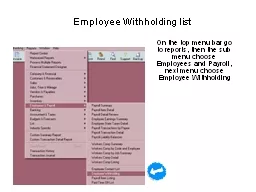
/
AIn the main menu at the top of the screen use the Views tab to sear AIn the main menu at the top of the screen use the Views tab to sear

AIn the main menu at the top of the screen use the Views tab to sear - PDF document

willow
willow . @willow
Follow
342 views
Uploaded On 2022-08-25

AIn the main menu at the top of the screen use the Views tab to sear - PPT Presentation

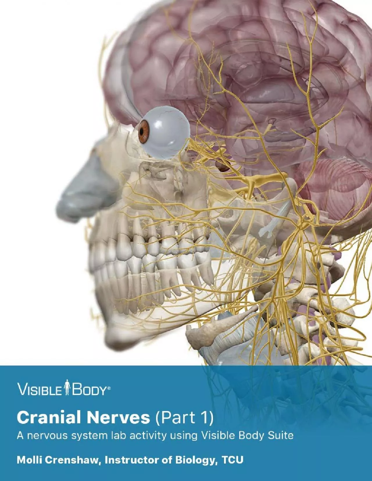
3 CerebellumMidbrain 7 Cranial nerve 07 VII 8 Cranial nerve 08 VIII 9 Cranial nerve 09 XI 10 Cranial nerve 10 X 11 Cranial nerve 11 XI 12 Cranial nerve 12 XII 4 Cerebral crus of the ID: 941637

nerve nerves select cranial nerves nerve cranial select brain optic branch fibers olfactory bone identify trigeminal system circle sensory

Share:

Link:

Embed:

Download Presentation from below link

Download Pdf The PPT/PDF document "AIn the main menu at the top of the scre..." is the property of its rightful owner. Permission is granted to download and print the materials on this web site for personal, non-commercial use only, and to display it on your personal computer provided you do not modify the materials and that you retain all copyright notices contained in the materials. By downloading content from our website, you accept the terms of this agreement.


Presentation Transcript

3 A.In the main menu at the top of the screen, use the Views tab to search by body systems. UnderNervous System Views, select 2. Brain, and make the following observations:1.Which two structures make up the central nervous system (CNS)?2.This view demonstrates the location of bones that form the cranium. What important roles does thecranium play in regard to the brain

?3.Identify each of the bones that form the cranium. Hide each of these bones after you’ve identifiedFrontalParietal (left)Temporal (left and right)Zygomatic (left and right)4.Rotate the image so that you are viewing the lateral surface of the left cerebral hemisphere. Selectand identify each of the following regions of the brain:Frontal lobe of the cerebrumPrece

ntral gyrus of the frontal cortexPostcentral gyrus of the parietal lobeParietal lobe of the cerebrumTemporal lobe of the cerebrumOccipital lobe of the cerebrum CerebellumMidbrain 7. Cranial nerve 07 (VII) 8. Cranial nerve 08 (VIII) 9. Cranial nerve 09 (XI) 10. Cranial nerve 10 (X) 11. Cranial nerve 11 (XI) 12. Cranial nerve 12 (XII) 4 Cerebral crus of the midbrainSup

erior colliculus of the midbrainInferior colliculus of the midbrainPons5. While it is not visible from this view, what part of the nervous system would extend below the 6. Select any of the cranial nerves in this view. Then, in the content box, select the caret at the top left. The caret will open a breadcrumb trail with a hierarchy of structures. In this hierarchy, s

elect "Cranial nerves" and use the definition to answer the following questions:a. How many pairs of cranial nerves are there in the human body?b. Cranial nerves are named for their connection to which part of the central nervous system?c. How many pairs of spinal nerves are there in the human body?B. In preparation for more extensive learning in lab, use Nervous Syst

em View 6. Cranial Nerves to identify the 12 pairs of cranial nerves. Be sure to use the Systems menu on the left side of the screen to deselect the skeletal, muscular, and digestive systems for the best view of the nerves. Use the space below to write out the scientific name of each nerve and a brief description of its 1. Cranial nerve 01 (I) 2. Cranial nerve 02 (II)

3. Cranial nerve 03 (III) 4. Cranial nerve 04 (IV) 5. Cranial nerve 05 (V) 6. Cranial nerve 06 (VI) 2 This lab activity is aligned with Visible Body’s Human Anatomy Atlas app.Learn more at visiblebody.com/professors We've split our Cranial Nerves lab activity into two parts. Part 1 is pre-lab exercises as well as exercises that incorporate cranial nerves I-VI.

Part 2 includes exercises covering cranial nerves VII-XII as well as post-lab exercises. 33 Trigeminal Nerves (IV) 32 Abducens Nerves (VI) 31 Trochlear Nerves (V) 30 Oculomotor Nerves (III) 29 Optic Nerves (II) 28 Olfactory Nerves (I) 27 Cranial Nerves 26 5. Brain 25 2. Brain 24 1. Brain 23 22 motor fibers terminate on which structures?d. As this branch transmits nerv

e impulses to and from the brain, which cranial foramen does it pass through? Which cranial bone is this foramen associated with?7. Based on your learning, how do you think damage to this nerve would present clinically?8. Click on the pathology icon and write out the symptoms of trigeminal neuralgia9. What types of tests would you devise to test the function of this n

erve in patients? 21 c. As this branch transmits nerve impulses toward the brain, which cranial foramen does it pass through? Which cranial bone is this foramen associated with?d. Which brain region do the fibers of this branch terminate on?5. Select the maxillary branch of the trigeminal nerve and read its description:a. This branch is sensory/motor/mixed (circle one

).b. These sensory fibers originate from receptors associated with which regions?c. Which foramen in the maxilla allows branches of this nerve to innervate the skin of the face?d. Which foramen in the zygomatic bone allows branches of this nerve to innervate the skin of the face?e. Select the and hide it. Observe the many branches off the maxillary branch of the trig

eminal nerve.f. As this branch transmits nerve impulses toward the brain, which cranial foramen does it pass through? Which cranial bone is this foramen associated with?g. Which brain region do the fibers of this branch terminate on?6. Select the mandibular branch of the trigeminal nerve and read its description:a. This branch is sensory/motor/mixed (circle one).b. Th

ese fibers originate from receptors associated with which regions? 20 E. Identification of the Trigeminal Nerves (V) Ophthalmic branchMandibular branchSemilunar ganglionMaxillaMaxillary branch 1. Select the region of the trigeminal nerve closest to the brain stem. How does its size compare to that of the other nerves you have studied?2. Each trigeminal nerve divides

into ______ major branches.3. Identify the 4. Select the ophthalmic branch of the trigeminal nerve and read its description:a. This branch is sensory/motor/mixed (circle one).b. These sensory fibers originate from receptors associated with which regions: c. Which extraocular muscle is innervated by each abducens nerve? What is its function?d. What is the function of

sensory fibers of the abducens nerves?4. What is the common purpose of the oculomotor, trochlear, and abducens cranial nerves?5. Based on your learning, how do you think damage to these nerves would present clinically?6. What types of tests would you devise to test the function of these nerves in patients? d.Be able to identify the following effector targets of the oc

ulomotor nerves, along with theirfunctions:Levator palpebrae superioris muscleSuperior rectus muscleMedial rectus muscleiv.Inferior rectus muscleInferior oblique musclee.Use the search bar to identify these additional targets of the oculomotor nerve. Write theirfunction in the space provided below:Ciliary musclesPupillary sphincter of the iris2.Select the trochlear ne

rves, and select Fade Others. Observe their location, and read theirdescription.a.Where do fibers of this nerve originate?b.Which foramina do the trochlear nerves pass through? Which cranial bone are these foraminaassociated with?c.Which extraocular muscle is innervated by each trochlear nerve? What is its function?3.Select the abducens nerves, and select Fade Others.

Observe their location, and read theirdescription.a.Where do fibers of this nerve originate?b.Which foramina do the abducens nerves pass through? Which cranial bone are these foraminaassociated with? 17 A. In Nervous System Views, select 6. Cranial Nerves. Hide the sphenoid, frontal, and temporal bones to best see the required structures. Select any part of the olfac

tory nerve and answer the following questions:1. Select the oculomotor nerves, and select Fade Others. Observe their location, and read their description.a. Are these nerves sensory/motor/mixed (circle one)?b. Where do fibers of this nerve originate?c. Which foramina do the oculomotor nerves pass through? Which cranial bone are these foramina associated with? Abducens

Nerves (VI)Superior orbital fissureLateral rectus muscle 16 Trochlear Nerves (IV)PonsCN 04 (IV) TrochlearSuperior oblique muscleSuperior orbital fissure 15 D. Identification of the Oculomotor Nerves (III), Trochlear Nerves (IV), and Abducens Nerves (VI) Oculomotor Nerves (III)CN 03 (III) OculomotorInferior oblique muscleInferior rectus muscleMedial rectus muscleLevat

or palpebrae superioris muscleSuperior rectus muscle e. Where is the optic chiasm located in relation to the hypothalamus?f. Posterior to the optic chiasm, fibers from the optic nerves travel in the form of optic _________ to the _____________.g. Based on the anatomy of the optic chiasm, do you think the right cerebral hemisphere will process vision from the right eye

, left eye, or both eyes?3. Based on your learning, how do you think damage to this nerve would present clinically?4. What types of tests would you devise to test the function of this nerve in patients? 1. In Nervous System Views, select 6. Cranial Nerves. Rotate the image so you are looking into the empty socket of the right eye. Select the optic nerve and then selec

t Fade Others. Observe its location and read the description.a. These nerves are sensory/motor/mixed (circle one).b. These nerves are responsible for the sense of _________________. c. They originate in the ______________________ of each eye. d. Which bone contains the optic foramina that the optic nerves pass through (be sure to identify this structure)?e. At which p

oint do the right and left optic nerves converge?f. Where do the optic tracts exist?2. Rotate the image so you are looking into the right cerebral hemisphere. Select the read the description.a. The optic chiasm is the crossing point of the optic nerves. Is it a partial or a complete crossing?b. The crossed fibers occupy the medial/lateral (circle one) part of the chia

sm.c. The uncrossed fibers occupy the medial/lateral (circle one) part of the chiasm.d. Where is the optic chiasm located in relation to the pituitary gland? 12 Optic Nerves (II) C. Identification of the Optic Nerves Optic chiasmPituitary glandCN 02 (II) OpticHypothalamus ii. The cribriform plate supports the ________________________ - the terminus of the ____________

__________________ - and is perforated by numerous ________________________ for the passage of the branches of the _____________________________. Olfactory bulbs of CN 01i. Are olfactory bulbs seated superior or inferior to the cribriform plate?ii. Zoom in to see the projections on the inferior surface of the bulbs. (Tip: You may need to hide the ethmoid bone to see t

hem well.)iii. Within each bulb, axons of olfactory _________ form synapses with dendrites and cell iv. If you are attempting to view the olfactory bulbs on a preserved specimen, they may be missing or incomplete. Why do you think this is?Olfactory tracts of CN 01 - Olfactory tracts are formed by axons/dendrites (circle one). Lateral stria of CN 01 - Follow this tract

to see which region of the brain it terminates in: Medial stria of CN 01 - Follow this tract to see which region of the brain it terminates in:f. Signals transmitted along the olfactory tracts terminate in the ________________________________area in the ___________________ lobe of the cerebral cortex.3. Based on your learning, how do you think damage to this nerve wo

uld present clinically?4. What types of tests would you devise to test the function of this nerve in patients? 1. In Nervous System Views, select 6. Cranial Nerves. Hide the sphenoid, frontal, and temporal bones to best see the required structures. Select any part of the olfactory nerve and answer the following questions:a. These nerves are sensory/motor/mixed (circle

one).b. These nerves are responsible for the sense of ________________________. c. Where do these nerves originate?2. Using this same view, identify the following structures and answer questions based on their descriptions. Cribriform plate of the ethmoid bonei. Fade the cribriform plate and observe its relationship with the olfactory nerves. Olfactory Nerves (I)La

teral stria of CN 01Medial stria of CN 01Olfactory bulbsOlfactory tracts B. Identification of the Olfactory Nerves (I) 9 Cranial Nerves CN 04 (IV) TrochlearCN 09 (IX) GlossopharyngealCN 05 (V) TrigeminalCN 10 (X) VagusCN 11 (XI) Spinal (Accessory)CN 01 (I) OlfactoryCN 03 (III) OculomotorCN 07 (VII) FacialCN 08 (VIII) VestibulocochlearCN 12 (XII) Hypoglossal 8 A. Begi

n by reviewing locations of major brain regions. Return to Nervous System Views and select 2. Brain, to help you identify the following structures on your specimen:1. Cerebrum2. Cerebellum3. Diencephalon4. Midbrain5. Pons6. Medulla oblongata 3. BrainCerebrumCerebellumPons 7 2. BrainCerebellumParietal lobePostcentral gyrus of parietal lobePonsFrontal lobePrecentral gyr

us of frontal cortex 6 Obtain a brain model or a preserved specimen. Use the following modules to guide your exploration of the brain and the cranial nerves. As you identify structures, use the book icon to access definitions so you can answer the questions below. You are responsible for the identification of all bolded terms. 1. BrainParietal boneTemporal boneZygomat