PPT-DIGESTION What is digestion?
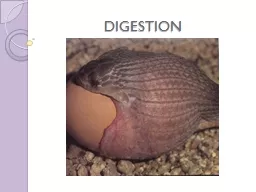
The process in which food is broken down into smaller molecules that can be absorbed into the body and are usable by cells Five Main Digestive Processes Ingestion
Download Presentation
"DIGESTION What is digestion?" is the property of its rightful owner. Permission is granted to download and print materials on this website for personal, non-commercial use only, provided you retain all copyright notices. By downloading content from our website, you accept the terms of this agreement.
Presentation Transcript
Transcript not available.