/
Life sciences Grade 11 CAPS Life sciences Grade 11 CAPS

Life sciences Grade 11 CAPS - PowerPoint Presentation

yoshiko-marsland
yoshiko-marsland . @yoshiko-marsland
Follow
356 views
Uploaded On 2019-12-11

Life sciences Grade 11 CAPS - PPT Presentation

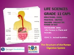
Life sciences Grade 11 CAPS structured clear practical Helping teachers unlock the power of NCS KNOWLEDGE AREA Life Process in Plants and Animals TOPIC 3 Animal Nutrition The Structure of the Human Digestive System ID: 770089

intestine food canal alimentary food intestine alimentary canal structure bile duct small liver called cells stomach question organs large

Share:

Link:

Embed:

Download Presentation from below link

Download Presentation The PPT/PDF document "Life sciences Grade 11 CAPS" is the property of its rightful owner. Permission is granted to download and print the materials on this web site for personal, non-commercial use only, and to display it on your personal computer provided you do not modify the materials and that you retain all copyright notices contained in the materials. By downloading content from our website, you accept the terms of this agreement.


Presentation Transcript

Life sciences Grade 11 CAPSstructured, clear, practical - Helping teachers unlock the power of NCS KNOWLEDGE AREA: Life Process in Plants and Animals.TOPIC 3: Animal Nutrition The Structure of the Human Digestive System

Summary of Presentation

Importance of Food Food is important because…It brings about growth by providing the substances that are required to make new cells .It serves as a source of energy for all living organisms. It is a source of regulating substances that are required for the functioning of the body. Some example of these substances are minerals and vitamins. These substances control and regulate the functions of the body . Food must include carbohydrates, fats, proteins, vitamins, mineral salts, fiber and water so that it can perform the above function.

Sources of foodMost organisms obtain their food from other living organisms.Can you think of any organism that does not need to do this? Why?

Sources of food Yes that’s right the organism is the green plant.Green plants you know are able to manufacture their own food by photosynthesis .Since green plants are able to make their own food they are called producers.All those organisms that are unable to make their own food are called consumers. The consumers can be divided into 3 groups based on what they eat. These 3 groups are herbivores, carnivores and omnivores .

Sources of foodThose organisms that feed on plant matter only are called herbivores.Those that feed on both plant and animal matter called omnivores .Those that feed on animal mater only are called carnivores.These feeding relationships can be shown by food chains . But remember in reality food chains do not exist in isolation.

Sources of foodIn any ecosystem, a number of food chains are interwoven.This is called a food web.This shows us that a particular organisms can belong to a number of different food chains.

Herbivores, carnivores & omnivoresAs seen in the previous slide the consumers have different diets.These organisms have many adaptations for their particular diets.We shall look at how the teeth of each group is adapted for the type of food the organism consumes.

Herbivores, carnivores & omnivores Herbivores:The incisors are broad and flattened for clipping of blades of grass.The molars and premolars are large and flat for grinding the grass.A gap occurs between the incisors and premolars to enable the tongue to move around during chewing . Dentition of a Herbivore

Herbivores, carnivores & omnivores This gap is called a diastema.Canines maybe absent as in the rabbit. If they are present they are usually short unless they are used for protection then they are longer. Molars are flattened.

Herbivores, carnivores & omnivores Carnivores:Here the incisors are short and pointed so that they can tear the food into small pieces that can be swallowed. Canines are long, pointed and curved to pierce and kill the prey.Molars are blade shaped, sharp and jaggered . Teeth of a Carnivore

Herbivores, carnivores & omnivores Omnivores:These organisms have long pointed canines These canines have a curved shape. The molars are blade shaped.These molars are sharp and or flattened.

Structure of the digestive system The digestive system is made up of 2 parts.These 2 parts are the alimentary canal and the associated organs. We shall look at each of these 2 parts in detail.Human digestive system with Associated organs

Structure of the alimentary canalThe alimentary canal is made up of the following parts: Mouth and mouth cavityPharynxOesophagusStomach Small intestine Large intestineAnus

Structure of the alimentary canal Mouth and Mouth Cavity:The mouth cavity is formed by the 2 lips in front .The tongue and teeth of the lower jaw forms the floor of the mouth cavity. The palate and teeth of the upper jaw forms the roof of the mouth cavity.The mouth cavity is lined by a type of mucous membrane that is made up of stratified epithelial connective tissue . Diagram showing mouth cavity

Structure of the alimentary canal Pharynx:The pharynx is the area where the nasal passage and the alimentary canal intersect each other .It occurs at the back of the mouth .Remember that the pharynx serves both the alimentary canal and the respiratory tract. Diagram showing the pharynx

Structure of the alimentary canal Therefore it leads to both the esophagus and the trachea.The trachea has an opening called the glottis.The pharynx leads to the respiratory tract via the glottis.The glottis has a covering called the epiglottis .

Structure of the alimentary canalWhen swallowing occurs, the glottis is closed by the epiglottis. Therefore the food cannot go down the trachea .This prevents choking. The Oesophagus:The oesophagus is a long muscular tube.It lies next to and behind the trachea . It leads from the pharynx to the stomach .

Structure of the alimentary canal The Stomach:The stomach is found in the abdominal cavity below the diaphragm.It is sac-like or bag-like organ .It has thick muscular walls.The opening between the stomach and oesophagus is closed by a valve . The position of the stomach

Structure of the alimentary canalThis valve is called cardiac sphincter.The opening between the stomach and small intestine is closed by another valve .This valve is called pyloric sphincter .The stomach

Structure of the alimentary canal The Small Intestine:This is a long muscular tube.It is about 2.5m to 4.5m long.The stomach leads into the small intestine .The small intestine is made up of 3 parts.These 3 parts are the duodenum, jejunum and ileum . Diagram showing the 3 parts of the Small Intestine

Structure of the alimentary canal The first part of the small intestine is called the duodenum. The hepato-pancreatic duct opens into the duodenum.This duct is formed when the common bile duct and the pancreatic duct join . The common bile duct comes from the gall bladder . The pancreatic duct comes from the pancreas .

Structure of the alimentary canal The jejunum is a short tube found between the duodenum and the ileum. Jejunum leads to the ileum.The ileum is the last part of the small intestine.It is also the longest part of the small intestine.It leads into the caecum . The caecum is the first part of the large intestine . The walls of the small intestine is made up of 4 layers .

Structure of the alimentary canal These four layers are:The outermost serosa,This is followed by a muscular layer. The muscular layer is made up of an outer layer of longitudinal muscles and an inner layer of circular muscles. T/s of wall of small intestine

Structure of the alimentary canal The muscular layer is followed by the sub mucosa.The sub mucosa is made up of connective tissue .The connective tissue includes blood vessels, lymph vessels and nerves.The sub mucosa is followed by the mucosa.The mucosa is also called the mucous membrane . It is the innermost layer . It is lined by columnar epithelium with goblet cells . The goblet cells secrete mucus .

Structure of the alimentary canal The mucous membrane has millions of tiny finger like projections.These finger like projections are called villi .The function of the villi is to increase the surface area for absorption of digested food.

Structure of the alimentary canal Each villus has the following structure:At the center of the villus there is large single lymph vessel called the lacteal .The lacteal is surrounded by connective tissue.A network of capillaries surround the lacteal . Structure of Villus

Structure of the alimentary canal These capillaries rejoin to form a venule.The venule leaves the villus. Then the entire villus is covered a single layer of columnar epithelial cells.Specialized cells called goblet cells are found between the epithelial cells .

Structure of the alimentary canal These goblet cells secrete mucus.Crypts of Lieberkuhn are found between the villi .Special compound glands are found between the villi of the duodenum.

Structure of the alimentary canal These glands are called the Brunner’s gland. Both the Crypts and Brunner gland secrete an alkaline mucus. The mucus has 2 functions:It serves as a lubricant, allowing for the easy movement of food along the intestine.It also protects the lining of the small intestine from the acid chyme coming from the stomach . The muscular walls of the small intestine enable peristalsis to occur. The peristalsis helps the food to moved along the small intestine . Peristalsis also ensures that the food is thoroughly mixed with the digestive juices .

Structure of the alimentary canal The Large IntestineThe ileum of the small intestine leads into the large intestine .The large intestine is made up of 3 parts. These 3 parts are the caecum, the colon and the rectum.The first part of the large intestine is called the caecum . It is a sac-like portion , into which the ileum opens . Parts of the large intestine

Structure of the alimentary canal The appendix is attached to the caecum. The colon is the second part of the large intestine.It is made up of 3 parts.These 3 parts are the ascending colon, transverse colon and the descending colon . The Colon

Structure of the alimentary canal The largest part of the large intestine is called the rectum .The rectum opens to the outside via the anus.

The associated organs :The organs that are associated with the alimentary canal are the: TongueTeethSalivary glandsPancreas Liver Gall bladder

The associated organs : The Tongue:Structure:The tongue is a movable muscular organ.It is attached at its back .Functions:Pushes the food between the teeth for chewing . It helps in mixing the food with saliva . It assists in forming the food into a small ball called the bolus . It helps with swallowing .

The associated organs : It also has taste buds. These taste buds serve as the organs of taste. The Teeth:There are 32 teeth in the adult.They are found embedded in the sockets in both the upper and lower jaw . There are 4 types of teeth . These are the: incisors, canines, premolars and molars . The dental formula tell us the number and type of teeth in 1 half of each jaw.

The associated organs :The human dental formulae is: 2.1.2.3 2.1.2.3According to the dental formula, one half of the upper jaw has 2 incisors, 1 canine, 2 premolars and 3 molars. From the formula we can see that the lower jaw has the same formula.If we want to calculate the total number of teeth in each jaw, then we multiply each row of the formula by 2 .

The associated organs :If we wish to calculate the total number of teeth found in the human then we multiply one row by 4. The incisors are chisel shaped. They are used for biting of pieces of food.The canines are conical shaped. The canines helps the incisors with tearing of pieces of food.The premolars are teeth with flat surfaces and they have 2 pointed cusps . They are used for grinding the food .

The associated organs :The molars are teeth with large flat surfaces with 3 to 5 pointed cusps. They are used for grinding and crushing the food. Human Teeth

The associated organs :The Salivary Glands: There are 3 pairs of salivary glands.They are found in the muscles around the mouth.These glands are also called exocrine glands because their secretions are carried by ducts.The salivary glands secrete salvia.

The associated organs : The Pancreas:The pancreas is a large gland.The pancreas is found in the loop of the duodenum .It is made up of two types of cells .These are the pancreatic cells and the islet of Langerhans.The pancreatic cells secretes pancreatic juice.The pancreatic juice is carried by the pancreatic duct . The pancreatic duct joins the common bile duct to form the hepato-pancreatic duct . Then it enters the duodenum .

The associated organs :The islet of Langerhans are specialized cells that are found amongst the pancreatic cells. The islet of Langerhans secretes 2 hormones .These 2 hormones are insulin and glucagon.Functions:The pancreas secrete pancreatic juice which contain digestive enzymes . It secretes the hormones insulin and glucagon which play a role in maintaining the blood sugar levels .

The associated organs : The Liver:This is a large organ.It is found in the abdominal cavity just below the diaphragm. It partially covers the stomach .It is made up of 2 lobes. These are the right and left lobe.The right lobe is larger than the left lobe. Position of the Liver

The associated organs : The liver is made up of cells called liver cells .The liver cells secrete a liquid called bile.The liver cells have tiny ducts called the hepatic duct . The bile is stored in the gall bladder . The cystic duct arises from the gall bladder . The hepatic duct joins the cystic duct to form the common bile duct . Structure of Liver

The associated organs :The common bile duct then goes onto join the pancreatic duct to form the hepato-pancreatic duct. The hepato-pancreatic duct opens into the duodenum. Functions of the liver:The liver has 3 general functions.These are storage, de-animation and detoxification.As a storage organ it converts excess glucose into glycogen and stores it . It also converts the excess glucose into fat and stores it . It also stores minerals such as iron and important vitamins such as vitamin A, D and B12 .

The associated organs :In is role in de-animation it breaks down the excess amino acids. This results in the formation of urea. The urea is excreted by the kidneys. The liver can detoxify some harmful substances and make them harmless.The liver secretes bile and the bile is stored in the gall bladder . Now lets look at the bile: Bile is a yellow green liquid . It is alkaline and has no enzymes . It is secreted by the liver cells . It is stored in the gall bladder .

The associated organs :When needed the bile is transported by the common bile duct.The common bile duct collects the bile from the liver cells and the gall bladder .The common bile duct then joins with the pancreatic duct to form the hepato-pancreatic duct.The hepato-pancreatic duct then carries the bile to the small intestine , where the bile has the following functions .

The associated organs : The water in the bile keeps the food fluid enabling easy movement along the alimentary canal .The bile salts neutralize the acid food coming from the stomach. They also break up the fats into tiny droplets. We say they emulsifies the fats. This increases the surface area for enzyme action . Bile salts help in the absorption of fats and fat soluble vitamins, D, E, A and K . Bile prevents the decomposition of food in the alimentary canal because it is an antiseptic .

Terminology : Producers: are organisms that are able to make their own food. Consumers: are organisms that are unable to make their own food.Herbivores : are those organisms that feed on plant matter only. Omnivores: are those organisms that feed on both plant and animal matter. Carnivores: are those organisms that feed on animal mater only.Food web : refers to a number of interwoven food chains.

Terminology : Diastema: is a gap that occurs between the incisors and premolars to enable the tongue to move around during chewing.Pharynx: is the area where the nasal passage and the alimentary canal intersect each other.Trachea: is the air tube that leads to the lungs. Glottis : is an opening of the trachea.Epiglottis: is the covering or lid of the glottis. Oesophagus: is a long muscular tube running from the pharynx to the stomach.Cardiac sphincter: is the valve between the oesophagus and stomach.

Terminology : Pyloric sphincter: is the valve found between the stomach and the caecum.Serosa: is the outermost layer of the wall of the small intestine.Villi: finger-like projections that line the innermost wall of the small intestine. Lacteal: the large lymph vessel found at the centre of villus. Crypts of Lieberkuhn: body found between the villi that secretes an alkaline mucus. Glands of Brunner: glands that occur between the villi that secrete an alkaline mucus.

Terminology :Bolus : a small ball of food that moves along the alimentary canal.Exocrine glands: these are glands whose secretions are carried by ducts. islet of Langerhans: specialized cells that occur in the pancreas and secrete the hormones insulin and glucagon. Bile: is a yellow green liquid secreted by liver cells in the liver. Gall bladder: structure in which bile is stored

Question 1 A large organ that is involved in de-animation, storage and detoxification is… A. Stomach B. Liver C. Pancreas D. Gall bladder

Question 2 Bile is secreted by the… A. Stomach B. Liver C. Pancreas D. Gall bladder

Question 3 Hormones that are involved in maintaining the blood sugar balance are secreted by the… A. Stomach B. Liver C. Pancreas D. Gall bladder

Question 4 The yellow green alkaline liquid is stored in the… A. Stomach B. Liver C. Pancreas D. Gall bladder

Question 5 The fluid that emulsifies fats is… A. Insulin B. Glucagon C. Bile D. None of the above

Question 6 The specialized cells that occur amongst the pancreatic cells in the pancreas is… A. Islet of Langerhans B. Liver cells C. Both A and B D. None of the above

Question 7 The conical shaped tooth that assist the incisors in tearing off food is the… A. Molars B. Premolars C. Incisors D. Canines

Question 8 The teeth that are used for biting of pieces of food is called… A. Molars B. Premolars C. Incisors D. Canines

Question 9 Large teeth that have 2 pointed cusps are called… A. Molars B. Premolars C. Incisors D. Canines

Question 10 The teeth with 3 to 5 cusps that are used for grinding and crushing the food is called… A. Molars B. Premolars C. Incisors D. Canines

Question 11 The number of teeth in the adult human is… A. 32 B. 24 C. 21 D. 36

Question 12 The glands that secrete saliva are called… A. Salivary glands B. Pancreas C. Liver D. None of the above

Question 13 An example of an exocrine gland… A. Salivary glands B. Pancreas C. Liver D. None of the above

Question 14 The caecum, colon and rectum are part of the… A. Small intestine B. Large intestine C. Stomach D. Oesophagus

Question 15 The duodenum, ileum and jejunum are a part of… A. Small intestine B. Large intestine C. Stomach D. Oesophagus

Question 16 Which layer of the wall of the small intestine is the connective tissue with blood vessels, lymph and nerves forms a part of? A. Serosa B. Muscular layer C. Sub mucosa D. Mucosa

Question 17 The outermost layer of the wall of the small intestine is the… A. Serosa B. Muscular layer C. Sub mucosa D. Mucosa

Question 18 The area where the alimentary canal and the respiratory tract cross each other is… A. Oesophagus B. Trachea C. Pharynx D. Peristalsis

Question 19 The narrow tube that runs from the pharynx to the stomach is the… A. Oesophagus B. Trachea C. Pharynx D. Peristalsis

Question 20 The cavity that is formed by the palate on top and the tongue at the bottom… A. Stomach B. Mouth C. Liver D. None of the above

Solutions to final assessment questions BBCDCA DC BA AAABACAC A B