/
Pigment Changes in Parsley Leaves during Storage in Controlled or Ethy Pigment Changes in Parsley Leaves during Storage in Controlled or Ethy

Pigment Changes in Parsley Leaves during Storage in Controlled or Ethy - PDF document

yoshiko-marsland
yoshiko-marsland . @yoshiko-marsland
Follow
406 views
Uploaded On 2017-03-02

Pigment Changes in Parsley Leaves during Storage in Controlled or Ethy - PPT Presentation

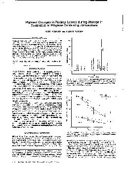
NAOKI YAMAUCHI and ALLEY E WATADA ABSTRACT Pigments were monitored in parsley leaves stored in air air 10 ppm GH or 10 O2 10 CO2 controlled atmosphere CA Chlorophylls a b as determined ID: 521245

NAOKI YAMAUCHI and ALLEY

Share:

Link:

Embed:

Download Presentation from below link

Download Pdf The PPT/PDF document "Pigment Changes in Parsley Leaves during..." is the property of its rightful owner. Permission is granted to download and print the materials on this web site for personal, non-commercial use only, and to display it on your personal computer provided you do not modify the materials and that you retain all copyright notices contained in the materials. By downloading content from our website, you accept the terms of this agreement.


Presentation Transcript

Pigment Changes in Parsley Leaves during Storage in Controlled or Ethylene Containing Atmosphere NAOKI YAMAUCHI and ALLEY E. WATADA ABSTRACT Pigments were monitored in parsley leaves stored in air, air + 10 ppm GH.,, or 10% O2 + 10% CO2 controlled atmosphere (CA). Chlorophylls a b, as determined with HPLC, decreased sharply in leaves held in air or air + 10 ppm GH4. YELLOWING of leafy vegetables, such parsley and spin- ach, occurs with degradation of chlorophyll (Chl). Tempera- Chl degradation may be due to differences in the degradative pathway, which is not clearly understood. Use of CA to retard color or Chl degradation has been shown with several vegetables including Brussels sprouts (Lyons and Rappaport, 1962), asparagus (Wang et al., 1971), and broccoli (Yang and Henze, 1988). Those studies showed that either green color or Chl content in the vegetable tissue was main- tained under controlled atmosphere storage, but they did not describe changes in the products of Chl degradation. Knowl- edge MATERIALS & METHODS FRESH ‘Forest Green’ parsley (Petrosefium crirum Nym.) was ob- tained from a local grower in Delaware and mature detached leaves free of defects or injury were used. About 150 of leaves were placed in a lightly covered 3.8L glass jar, and Author Watada is with the USDA-ARS Horticultural Crops &al- ity Laboratory, Beltsville Agricultural Research Center, Belts- ville, MD 20705. Author Yamauchis current address: Himeji College of Hyogo, Shinzaike-honcho, Himeji, Hyogo 670 Japan. 0 I I~..~...~~~~.........~....~I 0 Time (minutes) Fig. 1. -HPLC chromatogram of pigments extracted from fresh parsley leaves. HPLC system described in text. Chl-chlorophyll, Chlide-chlorophyllide, Phy- pheophytin, Phb-pheophorbide. 200 -Air --- Ethylene f.* 0 Days in Storage Fig. Z.-Changes in content of chlorophylls of parsley leaves stored in air with without 10 ppm Cti4 controlled atmo- sphere of 10% 0, 10% CO2 and 80% N,, as recommended by Apeland (1971). The gases were metered at a rate to maintain respiratory CO2 levels at about 0.5%. Sublots of leaves were removed for analysis after 0, 3 darkness. Aliquots 616-JOURNAL OF FOOD SCIENCE-Volume 58, No. 3, 1993 10000 8000 “.3.. *. . 8000 . # t - Ethylene . CA 00 0 2 Days in Storage Fig. 3. -Relative changes of chlorophyll a (A), chlorophyll a-l (B), and chlorophyllide (C) of parsley leaves held in air with or without 10 ppm C& or controlled atmosphere of 10% 0, 10% co, were for spectrophotometric (Shimadzu, Model W-260) analy- sis or passed through a Millipore filter (0.45 pm pore size) for HPLC analysis. tie HPLC with photodiode array detector system reported previ- ouslv (Yamauchi and Watada. 19911 was modified. The absorntion specira‘ of the pigments were recorded between 200 rim at the rate of 12 spectra/min. Pigments were separated by a Vydac C, ultrasphere column, 4.6 x 250 mm, using two solvents: A, 80% methanol, and B, ethyl acetate. Ethyl acetate was added to 80% meth- anol at a linear rate until a mixture was attained at the end of 20 min. The 5050 mixture then was run for an additional 20 min. Flow rate was 1 mUmin and injection volume was 50 pL. Identification of individual pigments from the acetone extract were carried out by methods described previously (Yamauchi and Watada, 1991). Chl &tent was determinid by the-method of Amon (1949); Tbe relative contents of lo-hvdroxvchloroohvll fCh1 a-1) and chlo- rophyllide were reported as peak area becauie iredared staidards were useful for peak identification, but for quantification. RESULTS HPLC of fresh parsley leaves showed sequential elution of neoxanthin, violaxanthin, lutein, Chl b, Chl a-l, Chl a p- carotene during a min run (Fig. 1). The elution time of chlorophyllide (Chlide) a b, pheophorbide a b, and pheophytin a are shown as reference points. Identity of each pigment was based on retention time, and in some in- stances were confirmed by the absorption spectra of the eluting peak. Chl a content in parsley leaves stored in air with or without GH., decreased at the same rate and were about 36% of the original level after 5 days storage (Fig. 2). Chl a of leaves exposed to air with or without &H, decreased at the same rate. Chl a of leaves held in CA decreased, but rate was slower and the content was maintained 20% longer than those in leaves held in air with or without GH,. Chl b decreased in all leaves and like Chl a, the decrease in samples held in air with or without &H, was greater than in those held in CA. The relative level of Chl a-l, the oxidized form of Chl a, was about 3% that Chl a, based on absorbance units (Fig. 3). Chl a-l of all treatments decreased by about 30% after one day of storage and then leveled off during the remainder of storage. The relative level of chlorophyllide was 10% of Chl a-l. With storage, a small accumulation was noted initially after storage in leaves from all treatments, but accumulation did not continue nor was it retained. The accumulation was re- tained longer in CA stored leaves where rate of Chl degradation was slower than that air or &H, treated samples. Pheophytin a content was low and decreased in leaves of all treatments (data not shown). The contents of xanthophylls, which included lutein, vio- laxanthin, and neoxanthin, decreased with yellowing of the leaves. However, at the same time, several new peaks occurred on the HPLC chromatograms with the yellowing (Fig. 4). The wavelengths of maximum absorbance of the new peaks were similar to the xanthophylls, such neoxanthin, vio- laxanthin and lutein (Table 1). Spectral properties of these peaks were also similar to parent xanthophyll, as shown for peaks 6,7 and compared with parent xanthophyll in Fig. Elution times of these new peaks (except peak 1) were considerably longer than those of the xanthophylls. DISCUSSION Chl CONTENT of parsley leaves decreased during storage at 20°C and was not hastened by 10 ppm CJ-I., which was un- expected. The effect of &H, may be apparent at a lower hold- ing temperature where the degradation of the control sample would not be so rapid. CA of 10% O2 and 10% CO, effective in reducing the rate of Chl degradation to the extent that the shelf life would be extended about 20% longer than that air-held samples. Wang (1979) postulated that COz inhibition of Chl degradation in broccoli may be to inhib- itory effect of CO, on &H, production or action. In examining the degraded products of Chl associated with yellowing of parsley leaves, Chl a-l had not accumulated whereas chlorophyllide a accumulated slightly, but to the extent of the amount of Chl degraded. In the orange flavedo, the peroxidase reaction has been shown to bleach chlorophyll (Huff, 1982). Thus the lack of chlorophyllide accumulation in the parsley may be to the presence of peroxidase reaction, which converted chlorophyllide to a colorless compound. The changes in the chlorophyllide were similar to those noted with spinach (Yamauchi and Watada, 1991), but were different from those of ethylene treated citrus. There chlorophyllase activity (Barmore, 1975; Shimokawa et al., 1978; Amir-Shapira et al., 1987) and chlorophyllide (Amir-Shapira et al., 1987) increased with yellowing. Neither the ethylene treatment nor CA had significant effect on formation of Chl a-l or chlorophyllide. With the decrease in the xanthophyll content, new pigments (peaks) were noted with the yellowing of the leaves. The new peaks were suspected to be esterified xanthophylls based on similar spectral characteristics and elution at a considerably later time than the xanthophylls. Esterified xanthophylls have been reported to appear with ripening of citrus flavedo (Eilati et al., 1972) and senescence of beech leaves (Tevini and Stein- maller, 1985). Others have reported that xanthophylls were esterified with fatty acids, such palmitic and linolenic acid (Eager and Schwenker, 1966). The esterified xanthophylls are Volume 58, No. 3, 1993-JOURNAL OF FOOD SCIENCE-617 PIGMENT CHANGES IN PARSLEY LEAVES. . 350 - 300 - 40- A 250 - 2o - El - lo- w Or 1% I 1 200 : 24 0% 150- !! % 100 - B d 50 - $8 I 100 % 0 r 0 Time (minutes) 300 Wavelength (nm) Fig. 5.-Absorption spectra of xanthophylls and new com- pounds that developed with yellowing of parsley leaves during storage. Fig. 4. - HPLC chromato- gram of pigments extracted from parsley leaves held in air with 10 ppm C& for 5 days at 20°C. Columns end solvent gradient of HPLC system described in text. 30 Table I-Spectral maxima of xanthophylls and new components that formed during storage of parsley leaves Xanthophylls and new components Wavelength (maxp Neoxanthin 413, 465 Violaxanthin 417, 469 Lutein 420, 475 Peak #1 417, 41, 471 Peak #2 421, 471 Peak #3 413, 465 Peak #4 415, 465 Peak #5 415, 465 Peak #6 413, 465 Peak #7 423. 473 Peak #6 Peak #9 421, 473 413, 465 ‘Wavelength (ma) of the pigments was measured by photodiode array detector. deposited in the plastoglobuli during senescence (Tevini and Steinmaller, 1985). In parsley leaves, the xanthophylls may be esterified with fatty acids and accumulated in the plasto- globuli of chloroplasts. These results imply that the pathway by which Chl is de- graded in parsley leaves was not altered by &H, or CA treat- ments. The lack of effect by &H, on the rate of degradation needs further study, in that others have reported it to hasten color and Chl degradation. Further study is also needed to determine if formation of xanthophyll pigments is interrelated with Chl degradation. With a better understanding of the effect of C;H, or CA individually (or in combination) on these pig- ment changes, the knowledge would be beneficial in devel- oping improved handling and storage conditions to maintain color quality of parsley and other leafy vegetables. Addition- ally, as are understood and enzymes that regulate the pathways are defined, the information would be useful in manipulating genes to develop cultivars that retain color and not senesce rapidly. REFERENCES Amir-Shapira., D., Goldschmidt, E.E., and Altman, A. 1967. Chlorophyll catabolism III senescing plant tissue: In viva breakdown intermediates -Continued on 618-JOURNAL OF FOOD SCIENCE-Volume 58, No. 3, 1993