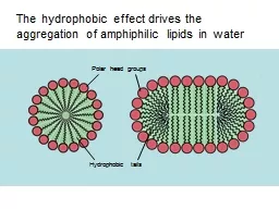
amphiphilic lipids in water Polar head groups Hydrophobic tails The shape of the lipid determines the structural features of the aggregate P acking at bilayer edges is crowded so bilayers merge their edges to form vesicles ID: 594139
Download Presentation The PPT/PDF document "The hydrophobic effect drives the aggreg..." is the property of its rightful owner. Permission is granted to download and print the materials on this web site for personal, non-commercial use only, and to display it on your personal computer provided you do not modify the materials and that you retain all copyright notices contained in the materials. By downloading content from our website, you accept the terms of this agreement.
Slide1
The hydrophobic effect drives the aggregation of
amphiphilic lipids in water
Polar head groups
Hydrophobic tailsSlide2
The shape of the lipid determines the structural features of the aggregateSlide3
P
acking at bilayer edges is crowded, so bilayers merge their edges to form vesicles
The bilayer separates the internal cavity from the external aqueous environment
bilayer
crowded edgesSlide4
Liposomes (vesicles) are bilayer-enclosed aqueous environments
leaflet
leaflet
bilayerSlide5
Biological membranes are heterogeneous lipid bilayers with proteinsSlide6
Protein and lipid content of biological membranes varies between speciesSlide7
Membrane lipid composition varies within a cellSlide8
Membrane
lipid composition
varies between leafletsSlide9
Membrane composition even varies within each leaflet! (non-random distribution)
Membrane microdomain (raft)Slide10
Atomic force microscopy reveals the presence of membrane microdomains (rafts)Slide11
Different lipid composition leads to different properties of the
membrane, like fluiditySlide12
Temperature also affects bilayer fluiditySlide13
At physiological temperatures, membrane bilayers are
(
and must be) quite fluidSlide14
E. coli can change its lipid composition to achieve ideal fluidity of its membraneSlide15
Lipids diffuse readily within one leaflet of the bilayer, but not between leafletsSlide16
Biological membranes are ‘fluid mosaics’ – proteins and lipids diffuse laterally
Photobleaching
allows measurement of
diffusion
rates in the membraneSlide17
The membrane ‘
skeleton,’ which shapes the cell, limits movement of proteins and lipidsSlide18
The ‘fencing-in’ and diffusion of lipids can be observed using fluorescent microscopy