PPT-HISTORY TMH Commissioned by the House of TATAs
Author : BabyDoll | Published Date : 2022-07-28
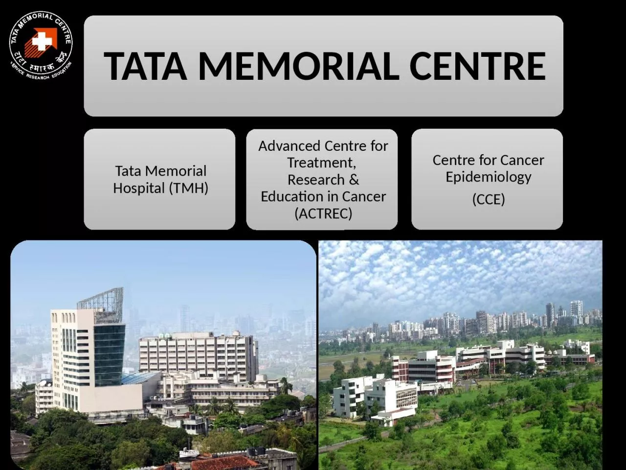
1941 CRI Commissioned 1952 TMH handed over to Ministry of Health 1957 TMH transferred under administrative control of DAE 1962 Tata Memorial Centre TMC TMH and
Presentation Embed Code
Download Presentation
Download Presentation The PPT/PDF document "HISTORY TMH Commissioned by the House of..." is the property of its rightful owner. Permission is granted to download and print the materials on this website for personal, non-commercial use only, and to display it on your personal computer provided you do not modify the materials and that you retain all copyright notices contained in the materials. By downloading content from our website, you accept the terms of this agreement.
HISTORY TMH Commissioned by the House of TATAs: Transcript
Download Rules Of Document
"HISTORY TMH Commissioned by the House of TATAs"The content belongs to its owner. You may download and print it for personal use, without modification, and keep all copyright notices. By downloading, you agree to these terms.
Related Documents