/
Bony thorax Muram  Ahmed Mohammed Bony thorax Muram  Ahmed Mohammed

Bony thorax Muram Ahmed Mohammed - PowerPoint Presentation

CitySlicker
CitySlicker . @CitySlicker
Follow
356 views
Uploaded On 2022-08-03

Bony thorax Muram Ahmed Mohammed - PPT Presentation

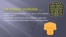
The Thoracic Cage Forms protective cage around heart lungs and other organs The skeleton of the Thorax consist of 1Sternum anteriorly Manubrium Body Gladiolus Xiphoid Process and costal cartilage ID: 933260

costal vertebrae typical ribs vertebrae costal ribs typical facets rib body articular vertebral transverse consist sacral curve thoracic process

Share:

Link:

Embed:

Download Presentation from below link

Download Presentation The PPT/PDF document "Bony thorax Muram Ahmed Mohammed" is the property of its rightful owner. Permission is granted to download and print the materials on this web site for personal, non-commercial use only, and to display it on your personal computer provided you do not modify the materials and that you retain all copyright notices contained in the materials. By downloading content from our website, you accept the terms of this agreement.


Presentation Transcript

Slide1

Bony thorax

Muram

Ahmed Mohammed

Slide2

The Thoracic Cage

Forms protective cage around heart, lungs, and other organs

Slide3

The skeleton of the Thorax consist of:-

1-Sternum(

anteriorly

)

Manubrium

, Body (Gladiolus), Xiphoid Process and costal cartilage 2-ribs(laterally)7 True Ribs5 False Ribs, 2 of them are floating ribs3-Clavicle(anterosuperior)4-Scapula(posterosuperior)5-Vertebrae(posteriorly)Cervical,Thoracic(dorsal), Lumbar, Sacral (Sacrum), Coccygeal (Coccyx)

Pg 14

Slide4

Slide5

1-The Sternum

It is flat bone with 2 surfaces, the anterior surface is rough while the posterior one is smooth. It articulates with the upper 7 ribs directly

anteriorly

via the costal cartilage.

Pg 120

Slide6

It is divided into 3 parts

:-

1-Manubrium

(2 inches in length) consist of:-

Jugular (sternal) notchArticulation with rib #1 & part of 2Clavicular Articular facets to form the sternoclavecular joint with the clavicleSternal Angle – where the 2nd rib articulates2-Body (Gladiolus)(4 inches in length) consist of:-Articulates w/ribs 2-6Xiphosternal joint

3-Xiphoid process

Articulate with the 7

th

cartilage

Slide7

2-The

Ribs

They

are 12 pairs

A-

7 True ribs-direct attachment to sternum called vertebrosternal attachment B- 5 False ribs -indirect or no attachment to sternum, they articulate with the sternum via the cartilage of the rob above (vertebrochondoral)Floating ribs

-make up 2 of 5 False ribs,

posteriorly

attached with the vertebral column but have no ventral

attachment

Pg 120

Slide8

Classification of ribs

1-According to sternum articulation:-

True , false, and

floaten

2-According to there shape:-

Typical ribs from 2ed to 9th A typical ribs number 1, 10, 11, 12

Slide9

Slide10

Slide11

Slide12

Anatomy of typical rib:-

Typical Rib is the rib that

articulates

with 2 vertebrae

bosteriorly

,each typical rib has 2 ends and shaft.1-Posterior end consist if:-Head, Neck ,Tubercle, and 2 articular surfaces in the head with crest in between.It artculates with the vertebrae with the same number, vertebral disc and the vertebrae above2-the anterior end is flat and articulates with costal cartilage3-the shaft consist of 2 surfaces(inner and outer), 2 borders( upper rounded in shape and lower sharp), Angle, and Subcostal Groove

Slide13

Typical Rib

Slide14

Anatomy of A typical rib:-

Atypical Ribs

#1-short, flat (S-I),and wide

#1, 10-12 articulate with only with the vertebrae with the same number so they have one

articular

surface in the head in the posterior end #11, 12 don’t articulate with transverse processes of the vertebrae , or anteriorly at allPg 121

Slide15

Atypical Ribs

Slide16

Typical Rib Articulation

Dorsal (P) Attachment Thoracic Vertebrae

Head of Rib

 2 costal facets

Superior costal facet

Inferior costal facet of vertebra above itIntervertebral disc Tubercle of Rib  Transverse Costal Facete.g. Rib #4 articulates with Superior Costal Facet and Transverse Costal Facet of T4 & Inferior Costal Facet of T3

Ventral (A) Attachment to Sternum

Via costal cartilage

Slide17

3-Vertebral

Column

Consist of 33 or 32 vertebrae

divided into

:-

1-Cervical 72-Thoracic 123-Lumbar 54-Sacrum (5 fused sacral vertebrae)5-Coccyx (4 or 3 fused coccygeal vertebrae)Extends from skull to pelvisSupports body, muscle attachmentVertebral CanalCreated by vertebral foramenContains + protects spinal cord

Pg 45, 18

Slide18

Normal vertebral curvatures:-

Cervical Region = Concave curve=

lordosis

Thoracic Region = Convex curve=

kyphosis

Lumbar Region = Concave curve=lordosisSacrum = Convex curve=kyphosisAbnormal CurvesScoliosis-abnormal lateral curve of more than 10° “twisted disease”Kyphosis-exaggerated thoracic curve “humped disease”Lordosis-accentuated lumbar curve “bent-backward disease”

Slide19

Slide20

Structure of typical vertebrae:-

1- Body

2-arch consist of 2

padicels

, 2

transvers processes, 1 spine, 2 superior and inferior articular process, 2 laminae, 2 superior and inferior vertebral notch which form the apophysial joint 3-intervertebral foramen between the body and the arch

Slide21

Slide22

A-Cervical

Vertebrae (7)

Classified into:-

1-A typical c/v:-#1,2,and 7

2-typical c/v:-#3-6

Slide23

Typical Cervical Vertebrae

**Transverse Foramen **

The body is small and oval

Superior

Articular

Facets face superoposteriorlyInferior Articular Facets face inferoanteriorlyAllows wide range of motionSpinous process fairly short, bifid (except for C7), point bake word Vertebral Foramen is Triangular Body is wider laterally than in A-P direction

Pg 31

Slide24

Slide25

Atypical Cervical Vertebrae

C1 – Atlas

No

body,has

2 arches (anterior and posterior) , 2

tuberosity (anterior and posterior) No Spinous ProcessSuperior Articular facets are kidney shapedC2 – AxisOdontoid Process = DensOther features typicalC7 has long and not

pifid

spine

Pg 32

Slide26

Thoracic Vertebrae (12)

**Transverse Costal facets directed

posterolateral

*S/I Costal facets on vertebral body*

Spinous

Processes long, point inferiorlySuperior Articular Facets face Dorsally/PosteriorlyInferior Articular Facets face Ventrally/AnteriorlyVertebral Foramen is CircularBody is Heart-shaped

Pg 119

Slide27

Slide28

A typical dorsal spine:-

Dorsal spine #1,10,11,and 12 has only one

Costal facets on vertebral body

Dorsal spine # 11 and 12 has no

Transverse Costal facets Other features are the same

Slide29

Lumbar Vertebrae (5)

Spinous

process is short, rectangular, thick, projects dorsally

Transverse process

Superior

Articular Facets face MediallyInferior Articular Facets face LaterallyVertebral Foramen is TriangularBody is large and Kidney-shapedPage 30

Slide30

Slide31

Sacrum & Coccyx

5 fused Sacral Vertebrae

Articulates with

1-5

th

Lumbar vertebra(lumbosacral joint)2-Coccyx(sacrococcygeal)3-Iliac bones(sacroiliac joint)Functions in weight transfer Has a body, sacral canal.Anteriorly consist of promontory , and ala

Sacrum

Slide32

Slide33

Sacrum

Posteriorly

it has 4 sacral foramina, 3 sacral crests(2 Lateral & median),sacral hiatus ,

inferolateral

angle, 2 corn

Slide34

Coccyx

3-4 fused

Coccygeal

vertebrae

Articulations

#1 articulates with 5th Sacral Vertebra (sacrococcygeal joint)Slightly different orientation in males vs. femalesNo canalHas transverse processes

Slide35

Intervertebral Discs

Absent between

C1 and C2

Sacrum and coccyx

The disc has 2 layers:-

1-Annulus FibrosusOuter collar of concentric rings Outer rings = ligamentsInner rings = fibrocartilage2-Nucleus PulposusInner discAct as Shock AbsorberPg 41