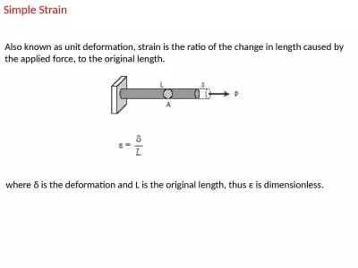
/
Maspin  Expression of Pancreatic Inflammation in an Obese Strain of Mice Maspin  Expression of Pancreatic Inflammation in an Obese Strain of Mice

Maspin Expression of Pancreatic Inflammation in an Obese Strain of Mice - PowerPoint Presentation

DancingDragonfly
DancingDragonfly . @DancingDragonfly
Follow
342 views
Uploaded On 2022-08-02

Maspin Expression of Pancreatic Inflammation in an Obese Strain of Mice - PPT Presentation

Janessa Pennington 1 Mariah Davis 1 Lindsey Haldiman 2 Alexey Glazyrin 2 Betty Herndon 1 Devika Kapuria 1 Agostino Molteni 1 1 UMKC School of Medicine 2 Truman Medical Center ID: 932740

maspin mice controls pancreas mice maspin pancreas controls liver exocrine stain treated cells inflammatory control amp endocrine ducts pancreatic

Share:

Link:

Embed:

Download Presentation from below link

Download Presentation The PPT/PDF document "Maspin Expression of Pancreatic Inflamm..." is the property of its rightful owner. Permission is granted to download and print the materials on this web site for personal, non-commercial use only, and to display it on your personal computer provided you do not modify the materials and that you retain all copyright notices contained in the materials. By downloading content from our website, you accept the terms of this agreement.


Presentation Transcript

Slide1

Maspin

Expression of Pancreatic Inflammation in an Obese Strain of Mice

Janessa Pennington1, Mariah Davis1, Lindsey Haldiman2, Alexey Glazyrin2, Betty Herndon1, Devika Kapuria1, Agostino Molteni11UMKC School of Medicine, 2Truman Medical Center

IntroductionPancreatic chronic inflammation often leads to glandular neoplasia. Up regulation of Maspin is a prognostic marker specific for this organ. This study evaluates pancreatic histopathological changes in a model of leptin knock out obese (ob/ob) mice to determine their expression of Maspin.

Ten mice (controls) received a normal diet and to ten more mice we added Ω3 (20% EPA, 50% DHA) to their food pellets, similar to an adult human dose of 4 grams per day.Two months later, at necropsy pancreases and livers were collected and stained with H&E.Liver glycogen determinations were made using image J method.

Anti-maspin stain therefore reflects the severe inflammatory reaction both in exocrine and endocrine pancreases of the Ω3 treated obese mice.Marked changes in glucose metabolism and severe diabetes despite the modest reduction of liver steatosis.

ReferencesBerardi et al, Role of Maspin in Cancer Clin & Translational Mwed 2:8, 2013Koutsounas I et al, World J, Gastroenterology 19:813-828, 2013

Half of the 10 Ω3 treated mice died during the two months of the study. The livers of controls showed diffuse steatosis reduced by 15-20% in the five Ω3 survivors. Anti-Maspin stain of controls showed few patchy areas of stain in the exocrine pancreas acini, some ducts and Langerhans islets. Stain was very intense in both the exocrine and endocrine pancreases of the Ω3 treated mice in the acini, ducts and peripancreatic fat and islets in particular. Blood glucose was very elevated (450 mg versus 171 mg in controls) with higher levels of insulin and increased insulin resistance. Liver glycogen determinations by image J method was 11.8% in the Ω3 group versus 17.55% of the controls.

Pancreas tissue of control (a) and Ω3 OB/OB mice (b

) 100x

The untreated exocrine pancreas showed scattered localized necrosis with destruction of the parenchyma and presence of acute inflammatory cells. Portions of the pancreatic ducts had the wall thickened with inflammatory cells mostly neutrophils in the parietal wall. Inflammation and larger lymph nodes were seen in the fat adjacent to the glandular areas where regular gland organization was partially destroyed with presence of necrotic cells and pyknotic nuclei.

Liver and pancreas control (a) and Ω3 OB/OB mice (b) 100x

a

b

Histopathology

Truman Medical Center and poster preparation by Mr. Joseph Moran, Office

of Marketing and Communication, UMKC SOM.

INTRODUCTION

METHODS

RESULTS

H&E STAINING LIVER AND PANCREAS

MASPIN STAINING- ENDOCRINE AND EXOCRINE PANCREAS

SUMMARY AND CONCLUSIONS

ACKNOWLEDGEMENTS

REFERENCES

a

a

b

b

a

a

b

b

b

Pancreas tissue of control (a) and Ω3 OB/OB mice (b) 400x