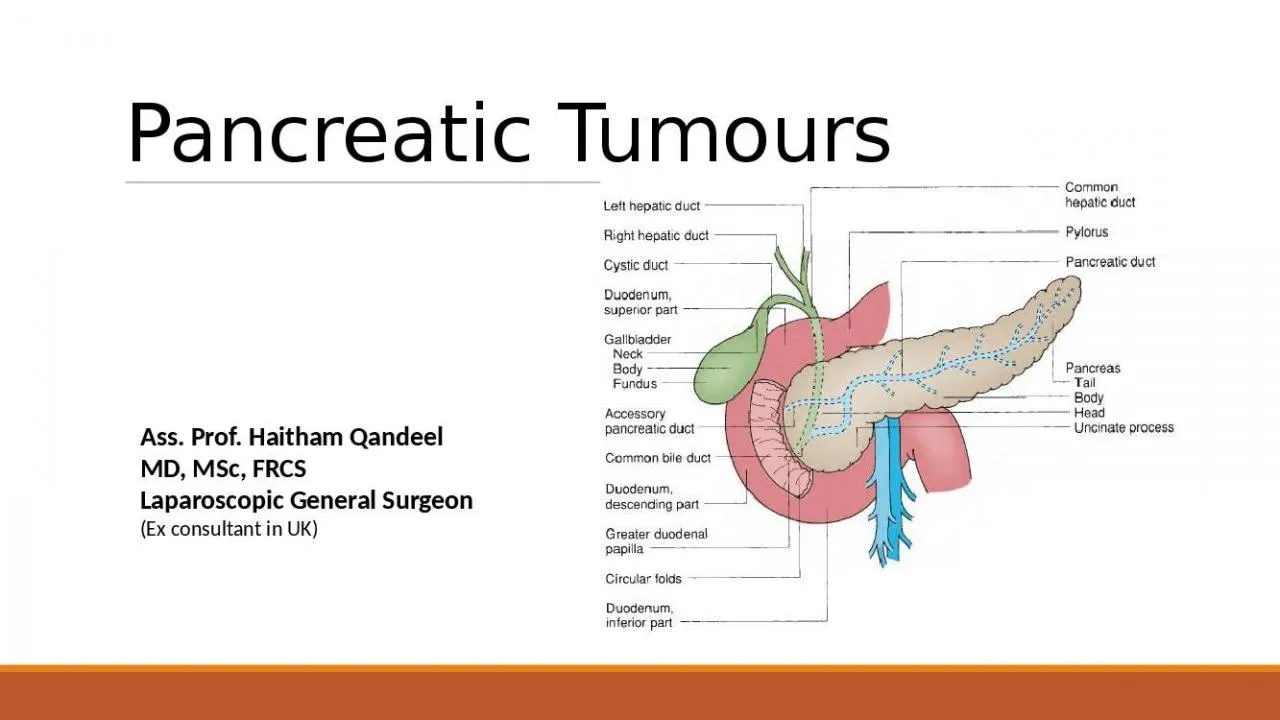
Prof Haitham Qandeel MD MSc FRCS Laparoscopic General Surgeon Ex consultant in UK STRUCTURE AND HISTOLOGY The pancreas has two major components the exocrine structure and the endocrine structure ID: 909514
Download Presentation The PPT/PDF document "Pancreatic Tumours Ass." is the property of its rightful owner. Permission is granted to download and print the materials on this web site for personal, non-commercial use only, and to display it on your personal computer provided you do not modify the materials and that you retain all copyright notices contained in the materials. By downloading content from our website, you accept the terms of this agreement.
Slide1
Pancreatic Tumours
Ass.
Prof.
Haitham
Qandeel
MD, MSc, FRCS
Laparoscopic General Surgeon
(Ex consultant
in
UK)
Slide2STRUCTURE AND
HISTOLOGYThe pancreas has two major components: the exocrine structure and the endocrine structure.
The exocrine structure of the pancreas forms 80-90% of the pancreatic mass, while the endocrine structure
forms
approximately 2%. The remainder of the pancreas is composed of extracellular matrix, blood vessels, lymph & nerves.The exocrine component secretes the enzymes responsible for digestion, and the endocrine component is critical in glucose homeostasis.
Slide3Exocrine Structure
The exocrine structure of the pancreas is composed of two main components: the acinar cells and the ductal network.
The acinar cells produce and secrete the enzymes responsible for digestion.
The role of the ductal system is to carry the digestive secretions to the duodenum.
The acinar cells are pyramidal cells with an apex that faces the pancreatic ductal network. Within the apex of the cells there are numerous zymogen granules, which contain the digestive enzymes for secretion into the ductal system.
20 - 40 acinar cells cluster together to form the functional unit called an acinus.
A second cell type in the acinus, the
centroacinar
cell, functions to secrete fluid and electrolytes of the correct pH into the pancreatic ductal system.
Histologic anatomy of the acinus.
A: Low-magnification view of a portion of the pancreas.
B: High-magnification view of a single acinus.
Slide4Endocrine Structure
The pancreatic islet cells are of neural crest origin and part of the family of
Amine
P
recursor Uptake
and
Decarboxylation
(APUD) cells.
The islets are composed of four cell types:Alpha (A) cells (10% of Islet cell mass) secrete glucagon, Beta (B) cells (70% of islet cell mass) secrete insulin and amylin, Delta (D) cells (5% of Islet cell mass) secrete somatostatin, and
F cells (15% of Islet cell mass) secrete pancreatic polypeptide.The distribution of endocrine cell types is not uniform throughout the pancreas:
B and D cells are uniformly distributed throughout the gland. A cells are concentrated in the body and tail of the pancreas. F cells are concentrated in the
uncinate
process.
This distribution is important clinically, since resection of different parts of the pancreas will have varying endocrine effects.
Diagram of a typical islet.
Slide5The most common of all
Simplified Classification of Pancreatic
Tumors
Slide6The most common of all
(IPMN) Important entity
Slide7Endocrine Tumors
Pancreatic Neuro-Endocrine Tumors
(
PanNETs, PETs, or PNETs) = islet cell tumors = pancreatic endocrine tumors: are neuroendocrine neoplasms that arise from cells of the endocrine and nervous system within the pancreas.Only 1 - 2% of clinically significant pancreas neoplasms are PanNETs.
Slide8Endocrine
Tumors
Most types of Pan NET are malignant (except
Insulinoma
). Generally, Pan NET (benign & malignant) can also be classified as functional or non-functional
.
Generally,
non-functional
PanNET are more common than functional.Functional PanNET (
insulinomas ”most common”, gastrinomas, Vasoactive
Intestinal Peptide (VIP)omas, glucagonomas
, and
somatostatinomas
).
Non-functional PanNET are not associated with a clinical syndrome, but can still produce symptoms related to the presence of the tumor or its metastases.
Immunohistochemistry can help identify the type of NET, as well as serve as a biomarker for diagnosis. For example, there is a unique occurrence of
psammoma
bodies in
somatostatinomas
(functioning
PanNET
) localized within the duodenum.
Slide9Endocrine
Tumors
Pan NETs are a type of neuroendocrine
tumor
(NET), representing about one third of GastroEnteroPancreatic
N
euroEndocrine
Tumors
(GEP-NETs). Aggressive PanNET
tumors have traditionally been termed "islet cell carcinoma“,
W
hile
others termed “islet cell
tumors
”.
Slide10Slide11CLASSIFICATION OF FUNCTIONAL PANCREATIC ENDOCRINE TUMORS
TUMOR (SYNDROME)
Arranged according to frequency
SOURCE
CLINICAL FEATURES
EXTRAPANCREATIC LOCATION
MALIGNANCY RATE
Insulinoma
Beta
cells
Whipple’s
Triad:
Hypoglycaemia
, CNS dysfunction
&
Reversal by glucose.
Rare
10%
Gastrinoma (Zollinger-Ellison)
G cells
Peptic ulcer
Diarrhea
Frequent
50%
VIPoma
(Verner-Morrison; WDHA; pancreatic cholera)
WDHA syndrome:
Watery
Diarrhea
,
Hypokalemia
,
& either
Achlorhydria
or
Acidosis.
10%
Most
Glucagonoma
Alpha cells
Diabetes, Dermatitis
(
necrolytic
migratory erythema)
Rare
Most
Somatostatinoma
Delta/S cells
Diabetes, Steatorrhea,
Gallstones
Rare
Most
Slide12Endocrine
TumorsInsulinomas
is the
most common type of Functional PanNET.90% of insulinomas are solitary,
90%
are sporadic, and
90%
are benign with location evenly distributed throughout the pancreas. 75% of gastrinomas are sporadic (25% are associated with Multiple Endocrine Neoplasia
type 1 MEN-1 syndrome), and all should be considered to be of malignant potential. Most gastrinomas are located in the gastrinoma
triangle and may be intrapancreatic, within the wall of the duodenum, or in a peripancreatic lymph node, and in most cases local resection (enucleation) may be adequate therapy.
Glucagonomas
usually present with a characteristic severe dermatitis (termed
necrolytic
migratory erythema) and are typically large and bulky and often with metastatic disease.
Slide13Pancreatic Cancer
Pancreatic cancer is the fourth leading cause of cancer death in USA, but in Jordan it is not one of the top five cancers.Currently, only 15-20% of patients diagnosed with pancreatic adenocarcinoma are candidates for pancreatic resection. 5-year survival is 15-20% for patients with resected disease and only 3% for all stages combined.
The nonspecific symptoms associated with early pancreatic cancer, the inaccessibility of the pancreas to examination, the aggressiveness of the
tumors
, and the technical difficulties associated with pancreatic surgery make pancreatic cancer one of the most challenging diseases treated by general surgeons. In recent years, significant advances have been made in our understanding of the pathogenesis and clinical management of pancreatic cancer.
Slide14RISK FACTORS FOR PANCREATIC CANCER
INCREASED RISK
POSSIBLE RISK
UNPROVED RISK
Demographic factors
Advancing age
Male sex
Black race
Geography
Socioeconomic status Migrant status
Host factors
Hereditary
Non-Polyposis
C
olorectal Cancer (HNPCC)
Familial breast cancer
Peutz-Jeghers
syndrome
Ataxia-telangiectasia
Familial
Atypical
M
ultiple
M
ole
M
elanoma (FAMMM)
Hereditary pancreatitis
Peptic ulcer surgery
Environmental factors
Tobacco
Diet Occupation
Alcohol Coffee
Radiation
Modified from Gold EB, Goldin SB. Epidemiology of and risk factors for pancreatic cancer.
Surg
Oncol
Clin
North Am
1998;7:67.
Slide15GENETIC ALTERATIONS IN PANCREATIC ADENOCARCINOMAS
GENE
CHROMOSOME LOCUS
FREQUENCY (%)
ONCOGENES
K-
ras
12
90
TUMOR-SUPPRESSOR GENES
p16
9p
95
p53
17p
50-75
DPC4
18q
55
BRCA2
13q
7
LKB1
17p
4
MKK4
19p
5
ALK4
12q
2
Genome maintenance 4 genes
bMSH2
,
hMLH1
2P, 3P
Reproduced with permission from
Hruban
RH. Pancreatic cancer: from genes to patient care.
J
Gastrointest
Surg
2001;5:583.
Slide16GENETIC SYNDROMES ASSOCIATED WITH HEREDITARY PANCREATIC CANCER
SYNDROME
MODE OF INHERITANCE
GENE
FOLD INCREASE IN RISK
MANIFESTATION OF PANCREATIC CANCER
Peutz-Jeghers
AD
STKll
140×
Hamartomatous polyps of the gastrointestinal tract; mucocutaneous melanin macules
Hereditary pancreatitis
AD
PRSSl
60×
Recurrent episodes of severe pancreatitis starting at a young age
Familial pancreatic cancer
Unknown
Unknown
18×
At least one pair of first-degree relatives with pancreatic cancer
FAMMM
AD
p16
20×
Multiple nevi, atypical nevi, melanomas
Familial breast cancer 2
AD
BRCA2
10×
Breast, ovarian, and pancreatic cancer
HNPCC
AD
MSH2HLHl
Unknown
Colonic, endometrial, and gastric cancers; mutator phenotype
AD, autosomal dominant; FAMMM, familial atypical multiple mole melanoma; HNPCC, hereditary nonpolyposis colorectal cancer.
Slide17SYMPTOMS
OF PANCREATIC CANCER
SYMPTOM
PATIENTS (%)
SYMPTOM
PATIENTS (%)
HEAD
BODY AND TAIL
Weight loss
92
Weight loss
100
Jaundice
82
Pain
87
Pain
72
Weakness
43
Anorexia
64
Nausea
43
Dark urine
63
Vomiting
37
Light stools
62
Anorexia
33
Nausea
45
Constipation
27
Vomiting
37 Hematemesis 17 Weakness 35 Melena 17 Pruritus 24 Jaundice 7 Diarrhea 18 Fever 7 Melena 12 Diarrhea 3 Constipation 11 Fever 11 Hematemesis 8
Slide18SIGNS
OF PANCREATIC CANCER
SIGN
PATIENTS (%)
SIGN
PATIENTS (%)
HEAD
BODY AND TAIL
Jaundice
87
Palpable liver
33
Palpable liver
83
Tenderness
27
Palpable gallbladder
29
Abdominal mass
23
Tenderness
26
Ascites
20
Ascites
14
Jaundice
13
Abdominal mass
13
Diarrhea
3
Slide19Presenting Signs and Symptoms of Pancreatic Tumours
Pancreatic cancer can occur anywhere in the pancreas, but it occurs in the pancreatic head in approximately 75% of cases.
Patients with cancer in the head of the pancreas often present with obstructive jaundice secondary to occlusion of the
intrapancreatic
bile duct.Patients with cancer in the body and tail of the pancreas often present with abdominal pain and other vague abdominal symptoms as these tumors do not obstruct the bile duct and lead to obvious clinical signs. As a result, tumors in the pancreatic head are often picked up at an earlier stage.Similarly, in benign pancreatic diseases such as chronic pancreatitis, disease in the pancreatic head may cause benign biliary strictures and jaundice, whereas disease in the body and tail more often presents with abdominal pain.
Patients with cancer in the pancreatic head often have invasion of the adjacent duodenum. They may present with or develop signs and symptoms of duodenal or gastric outlet obstruction requiring
gastrojejunostomy
.
In addition, as pancreatic cancer progresses, the nervous plexuses along the celiac axis in the retroperitoneum can be invaded by tumor, causing the characteristic intractable back pain. Celiac ganglion blockade (sympathectomy
) or neurolysis using alcohol can provide significant pain relief by interrupting these somatic fibers.
Slide20Investigations
The best imaging technique for a pancreatic neoplasm is
Computed
T
omography (CT) scan - pancreatic
protocol.
MRCP offers no significant advantages over CT.
MRI
can be considered an alternative in patients with allergies to iodinated contrast agents and in patients with renal insufficiency.MRCP with secretin stimulation provides a clearer view of the ductal system and of its relations with cystic lesion of the pancreas, thus allowing better diagnostic classification of the IPMN.
Endoscopic ultrasonography (+/- biopsy) is particularly useful in localizing tumors in patients with
gastrinoma and insulinoma.ERCP may add brush cytology in some cases, but very little
yeald
.
ERCP
may be useful for palliative biliary stent insertion.
Slide21Investigations
Computed tomography scan is the preferred non-invasive imaging test for the diagnosis and staging of pancreatic cancer demonstrating the primary lesion and its relationship to adjacent visceral vessels as well as metastatic disease to the liver and peritoneum.
CT-scan abdomen of a patient with adenocarcinoma of the pancreas.
A:
The obstructed and dilated common bile duct (light arrow) and pancreatic duct (dark arrow) can be seen.
In the adjacent cross section
(B)
, a large mass is present in the head of the pancreas (arrow).
Slide22Investigations
The strengths of Endoscopic ultrasonogram (EUS):Clarification of small lesions (<2 cm) when CT findings are questionable or negative,
Detection of malignant lymphadenopathy,
Detection of vascular involvement, and
Ability to perform EUS-guided fine-needle aspiration (FNA) for definitive diagnosis and staging. EUS is not effective in assessing metastatic disease to the liver.EUS of a 2.2-cm mass in the head of the pancreas. The transducer tip is located in the duodenum. The dilated common bile duct and gallbladder (GB) can be seen at the top of the image. The pancreatic duct (PD) is also dilated. The mass involves the portal vein (PV).
Slide23Investigations
Endoscopic retrograde cholangiopancreatography in a patient with adenocarcinoma of the pancreas demonstrates a stricture of both the distal common bile duct and the pancreatic duct (arrow).
Although ERCP is reliable in confirming the presence of a clinically suspected pancreatic cancer, it should not be used routinely.
Diagnostic ERCP should be reserved for patients with presumed pancreatic cancer and obstructive jaundice in whom no mass is demonstrated on CT, symptomatic but
nonjaundiced
patients without an obvious pancreatic mass, and patients with chronic pancreatitis who develop jaundice.
Slide24Laboratory Tests
In patients with cancer of the head of the pancreas, an increase in serum total bilirubin, alkaline phosphatase, and γ-glutamyl
transferase, indicating
bile duct obstruction
. In patients with localized cancer of the body and tail of the pancreas, laboratory values are frequently normal early in the course. Patients with pancreatic cancer may also demonstrate a normochromic anemia and hypoalbuminemia
secondary to the nutritional consequences of the disease.
In patients with jaundice, the
Prothrombin
Time (PT) can be abnormally prolonged. This usually is an indication of biliary obstruction, which prevents bile from entering the gastrointestinal tract and leads to malabsorption of fat-soluble vitamins and decreased hepatic production of vitamin K-dependent clotting factors. The prothrombin time can usually be normalized by the administration of
parenteral vitamin K. Serum amylase and lipase levels are usually normal in patients with pancreatic cancer
Slide25Laboratory Tests
The most extensively studied tumor marker is CA 19-9, a Lewis blood group-related mucin glycoprotein.
Approximately
5%
of the population lacks the Lewis gene and therefore cannot produce CA 19-9. The accuracy of the CA 19-9 level in identifying patients with pancreatic adenocarcinoma is only about 80-85%. The combined use of CA 19-9 and CT, EUS, or ERCP can improve the accuracy of the individual tests, so that the combined accuracy approaches 100% for the diagnosis of pancreatic cancer. Levels of CA 19-9 correlates with prognosis and tumor recurrence.
Slide26Laboratory Tests
In general, higher CA 19-9 values before surgery indicate an increased size of the primary tumor and increased rate of unresectability.
In addition, the CA 19-9 level has been used to
monitor
the results of neoadjuvant and adjuvant chemoradiation therapy in patients. Increasing CA 19-9 levels usually indicate recurrence or progression of disease, whereas stable or declining levels indicate a stable tumor burden, absence of recurrence on imaging studies, and an improved prognosis.
Slide27Pancreaticoduodenectomy
(
whipple’s
procedure or modified methods) for
tumors
in the Head, Neck, or
Uncinate
Process of pancreas.
Distal pancreatectomy for
tumors in the body and tail of pancreas.
Treatment
Slide28COMPLICATIONS AFTER PANCREATICODUODENECTOMY
COMMON
Delayed gastric emptying
Pancreatic fistula
Intra-abdominal abscess
Hemorrhage
Wound infection
Metabolic
Diabetes
Pancreatic exocrine insufficiency
UNCOMMON
Fistula
Biliary
Duodenal
Gastric
Organ failure
Cardiac
Hepatic
Pulmonary
Renal
Pancreatitis
Marginal ulceration
From Yeo CJ, Cameron JL. Pancreatic cancer.
Curr
Probl
Surg
1999;36:61, with permission.
Slide29Treatment
Perioperative mortality rates following
pancreaticoduodenectomy
have fallen to the range of 2% to 5%, although perioperative complications occur in approximately 40% of patients.
The survival rate after pancreaticoduodenectomy for pancreatic cancer is approximately 20%, with factors influencing survival including
tumor
size, margin status, and node status.
Most
data support the role for adjuvant therapy, either chemotherapy or chemoradiation
, for patients following resection of pancreatic cancer. Surgical palliation of patients with pancreatic cancer located in the head found to be
unresectable at laparotomy includes biliary stents, biliary bypass, gastrojejunostomy
, and chemical
splanchnicectomy
to palliate the symptoms of jaundice, duodenal obstruction, and pain, respectively.
Slide30THANK
YOU