PDF-(READ)-Anatomy in Diagnostic Imaging
Author : DeannaOconnell | Published Date : 2022-09-04
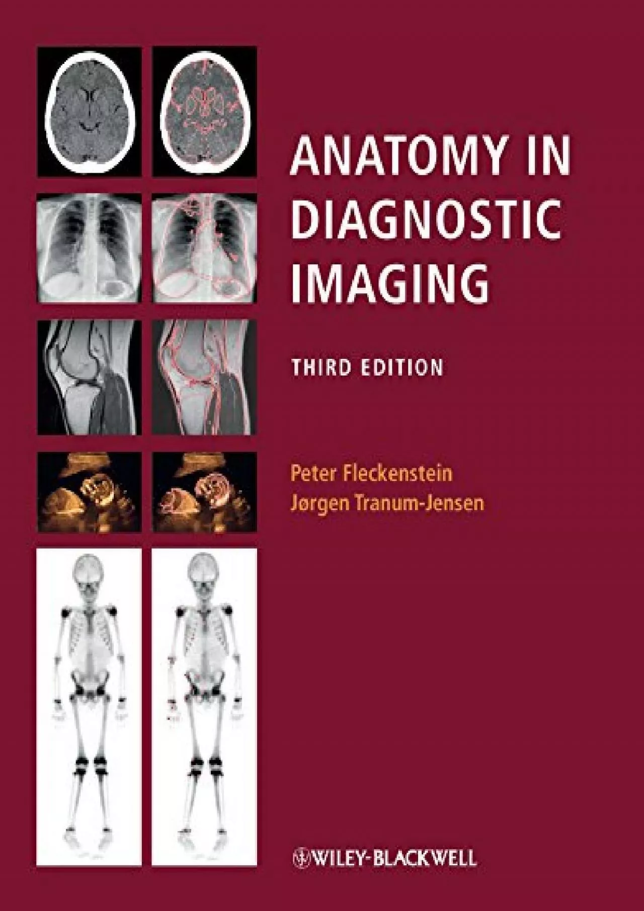
Now in its third edition Anatomy in Diagnostic Imaging is an unrivalled atlas of anatomy applied to diagnostic imaging The book covers the entire human body and
Presentation Embed Code
Download Presentation
Download Presentation The PPT/PDF document "(READ)-Anatomy in Diagnostic Imaging" is the property of its rightful owner. Permission is granted to download and print the materials on this website for personal, non-commercial use only, and to display it on your personal computer provided you do not modify the materials and that you retain all copyright notices contained in the materials. By downloading content from our website, you accept the terms of this agreement.
(READ)-Anatomy in Diagnostic Imaging: Transcript
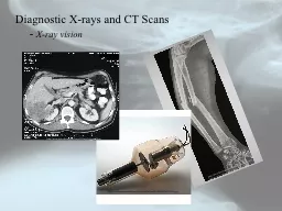
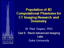
Now in its third edition Anatomy in Diagnostic Imaging is an unrivalled atlas of anatomy applied to diagnostic imaging The book covers the entire human body and employs all the imaging modalities used in clinical practice xray CT MR PET ultrasound and scintigraphy An introductory chapter explains succinctly the essentials of the imaging and examination techniques drawing on the latest technical developmentsIn view of the great strides that have been made in this area recently all chapters have been thoroughly revised in this third edition The books original and didactically convincing presentation has been enhanced with over 250 new images There are now more than 900 images all carefully selected in order to be userfriendly and easytoread due to their high quality and the comprehensive anatomical interpretation directly placed alongside every oneBoth for medical students and practising doctors Anatomy in Diagnostic Imaging Third edition will serve as the goto allround reference collection linking anatomy and modern diagnostic imaging. Collection, Packaging, Shipping. Overview. Sample Collection. Just In Time Training. Diagnostic Sampling: Overview. Before You Begin. Test specifications from laboratory. Samples needed, any special media . - . X-ray vision. http://www.bjwinslow.com/albums/medicalcharts/broken_arm_radius_and_ulna_x_ray_10.jpg. http://www.museumboerhaave.nl/AAcollection/AAJPEGS/M22/9955.jpg. http://www.uab.edu/surgonc/cases/GI/case2/ctscanof.htm. W. Paul Segars, PhD. Carl E. Ravin Advanced Imaging Labs. Duke University. Myocardial SPECT. Digital phantom. Emission Computed Tomography (ECT). X-ray CT. Transmission Computed Tomography (TCT). Medical Imaging Simulation. New diagnostic tests been studied extensively. Some 90% of immunogenicity. However, even fully therapeutic antibodies has been very successful. Well-known examples of are applied for in ammatory disea Gregg C. Daversa. Director, Business Development. West . Physics. Objectives. Identify which JC-accredited facilities will be impacted by the 2014 standard changes, and the dates for full compliance. Tim Shoen, MD. Campaign for Quality. October 17, 2014. Disclosure. No financial interest to disclose. Thanks to Mark Graber, MD, President, SIDM.. Sue Sheridan. Wall Street Journal. The Biggest Mistake Doctors Make. 2018. Committee on Acute Care Surgery, Canadian Association of General Surgeons. DIAGNOSTIC IMAGING MODALITIES. 4. Melissa Hanson MD, and. . Jacinthe. . Lampron. MD. Committee . on Acute Care Surgery, Canadian Association of General Surgeons. BRACHIAL PLEXUS IMAGING :. Basic anatomy. Pathologies affecting Brachial Plexus. Modalities of Imaging :. Conventional methods.. Ultrasound. CT and CT . Myelography. MR. ANATOMY : FORMATION : . Derived from anterior primary rami of C5 to C8 and T1.. structural . organisation. Anatome. (. ana. =up. . tomy. =cut) . Anatomise. Dissection. Practical applied science which forms firm foundation of art of healing(medicine). May 2019. Available Diagnostic Technologies. 2. Disease Diagnostics. Page. Ancsin:. . 15-T-073. Companion Assays for the Diagnosis of Deficiencies in Triglyceride Clearance for the Treatment of Hypertriglyceridemia (HTG). By: . Dr. Ammar Ismail. Introduction:. Anatomy. :( Greek word). Ana -------- a part. Tomy. ------- cut . Anatomy: . is the study of the structures of a body and relation of its parts. The subject is usually studied by dissection and observation.. WHAT IS EVIDENCE - BASED MEDICINE (EBM)?. The translation of medical research into clinical practice. Integration of best research evidence with clinical experience and patient values. Knowing how to use clinical literature to ensure optimal patient care. – An Introduction – . Dr . Rudra. . Pratap. . Pandey. Prof. VSR. Conventional radiography. Digital radiography (CR & DR). A. Equipment details. B. Safety issues – Radiation . Advantages . . radiologi. IPSG.1 – . Identifikasi. . pasien. . secara. . benar. IPSG.2 – . Melaporkan. . hasil. . pemeriksaan. yang . kritis. , . serah. . terima. . pasien. ACC.2.2.1 – . Radiologi.
Download Document
Here is the link to download the presentation.
"(READ)-Anatomy in Diagnostic Imaging"The content belongs to its owner. You may download and print it for personal use, without modification, and keep all copyright notices. By downloading, you agree to these terms.
Related Documents