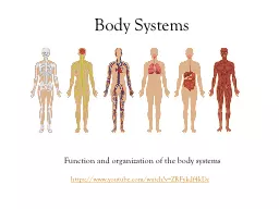
/
Concepts of Biology: The Body’s Systems Concepts of Biology: The Body’s Systems

Concepts of Biology: The Body’s Systems - PowerPoint Presentation

FluffyFace
FluffyFace . @FluffyFace
Follow
342 views
Uploaded On 2022-07-28

Concepts of Biology: The Body’s Systems - PPT Presentation

An arctic fox is a complex animal well adapted to its environment Credit Keith Morehouse USFWS CONCEPT IN ACTION Watch this Discovery Channel video on thermoregulation to see illustrations of the process in a variety of animals ID: 931046

system credit work modification credit system modification work blood muscle action body www concept heart http watch cells food

Share:

Link:

Embed:

Download Presentation from below link

Download Presentation The PPT/PDF document "Concepts of Biology: The Body’s System..." is the property of its rightful owner. Permission is granted to download and print the materials on this web site for personal, non-commercial use only, and to display it on your personal computer provided you do not modify the materials and that you retain all copyright notices contained in the materials. By downloading content from our website, you accept the terms of this agreement.


Presentation Transcript

Slide1

Concepts of Biology:

The Body’s Systems

Slide2

An arctic fox is a complex animal, well adapted to its environment.

(Credit

:

Keith

Morehouse

, USFWS)

Slide3

CONCEPT IN ACTION

Watch this

Discovery Channel video

on thermoregulation to see illustrations of the process in a variety of animals

.

https://www.youtube.com/watch?v=

NJEBfl_LKno

Slide4

The body is able to regulate temperature in response to signals from the

nervous system

.

Slide5

The human excretory system is made up of the kidneys, ureter, urinary

bladder, and urethra

. The kidneys filter blood and form urine, which is stored in the bladder until it is

eliminated through

the urethra. On the right, the internal structure of the kidney is shown.

(Credit

:

modification of

work by NCI, NIH)

Slide6

The components of the human digestive system

Slide7

Digestion of food begins in the mouth.

Food

is masticated by teeth and

moistened by

saliva secreted from the salivary

glands. Enzymes

in the saliva begin to digest starches and

fats. With

the help of the tongue, the resulting bolus is moved into the esophagus by

swallowing.

(Credit:

modification

of work by Mariana Ruiz

Villareal

)

Slide8

The large intestine reabsorbs water from undigested food and stores waste until it

is eliminated

.

(Credit

: modification of work by Mariana Ruiz

Villareal

)

Slide9

The stomach has an extremely acidic environment where most of the protein

gets digested

.

(Credit

: modification of work by Mariana Ruiz

Villareal

)

Slide10

For humans, a balanced diet includes fruits, vegetables, grains, protein, and

dairy.

(Credit: USDA)

Slide11

CONCEPT IN ACTION

Explore

this

interactive U.S.

Department of Agriculture website

to learn more about each food group and the recommended daily amounts

.

http://www.choosemyplate.gov/food-groups

/

Slide12

Air enters the respiratory system through the nasal cavity, and then passes through the pharynx and the trachea into the lungs. (Credit: modification of work by NCI)

Slide13

CONCEPT IN ACTION

Watch this

video

for a review of the respiratory system

.

https://www.khanacademy.org/science/health-and-medicine/respiratory-system/Respiratory-system-introduction/v/the-lungs-and-pulmonary-

system

Slide14

The heart is divided into four

chambers:

two

atria

and two ventricles.

Each chamber

is separated by one-way valves. The right side of the heart receives

deoxygenated blood

from the body and pumps it to the lungs. The left side of the heart pumps blood to the

rest of

the body.

Slide15

In each cardiac cycle, a series of contractions (systoles) and relaxations (diastoles

) pumps

blood through the heart and through the body

.

During cardiac diastole, blood flows into the heart while all chambers are relaxed.

Then

the ventricles remain relaxed while atrial

systole pushes

blood into the

ventricles.

Once

the atria relax again, ventricle systole pushes blood out

of the

heart.

Slide16

CONCEPT IN ACTION

Visit the

this

website

to see the heart’s pacemaker, or electrocardiogram system, in action

.

http://www.nhlbi.nih.gov/health/health-topics/topics/hhw/

electrical

Slide17

The arteries of the body, indicated in red, start at the aortic arch and branch to supply the organs and muscles of the body with oxygenated blood. The veins of the body, indicated in blue, return blood to the heart. The pulmonary arteries are blue to reflect the fact that they are deoxygenated, and the pulmonary veins are red to reflect that they are oxygenated. (Credit: modification of work by Mariana Ruiz

Villareal

)

Slide18

The pituitary gland sits at the base of the brain, just above the brain stem

.

The parathyroid

glands are located on the posterior of the thyroid gland

.

The

adrenal glands are

on top

of the

kidneys.

The pancreas is found between the stomach and the small intestine.

(Credit:

modification

of work by NCI, NIH)

Slide19

The anterior pituitary stimulates the thyroid gland to release thyroid hormones T

3

and T

4

. Increasing levels of these hormones in the blood result in feedback to the hypothalamus and anterior pituitary to inhibit further signaling to the thyroid gland. (Credit: modification of work

by Mikael Häggström)

Slide20

CONCEPT IN ACTION

Explore the human skeleton by viewing the following

video

with digital 3D sculpturing

.

https://www.youtube.com/watch?v=

7vLAE_P_u_4

Slide21

The axial skeleton, shown in blue, consists of the bones of the skull,

ossicles

of the middle ear, hyoid bone, vertebral column, and thoracic cage. The appendicular skeleton, shown in red, consists of the bones of the pectoral limbs, pectoral girdle, pelvic limb, and pelvic girdle. (Credit: modification of work by Mariana Ruiz

Villareal

)

Slide22

Sutures are fibrous joints found only in the skull

.

Cartilaginous

joints are

bones connected

by cartilage, such as between

vertebrae.

Synovial

joints are the only joints that

have a

space or “synovial cavity” in the joint.

Slide23

The body contains three types of muscle tissue: skeletal muscle, smooth muscle,

and cardiac

muscle. Notice that skeletal muscle cells are long and cylindrical, they have multiple nuclei

, and

the small, dark nuclei are pushed to the periphery of the cell. Smooth muscle cells are short

, tapered

at each end, and have only one nucleus each. Cardiac muscle cells are also cylindrical,

but short

. The cytoplasm may branch, and they have one or two nuclei in the center of the cell.

(Credit: modification

of work by NCI, NIH; scale-bar data from Matt Russell)

Slide24

A skeletal muscle fiber is surrounded by a plasma membrane called the sarcolemma

, with

a cytoplasm called the sarcoplasm. A muscle fiber is composed of many fibrils packaged

into orderly

units. The orderly arrangement of the proteins in each unit, shown as red and blue lines

, gives

the cell its striated appearance.

Slide25

CONCEPT IN ACTION

View this

animation

to see how muscle fibers are organized

.

https://www.youtube.com/watch?v=

XoP1diaXVCI

Slide26

Neurons contain organelles common to other cells, such as a nucleus

and mitochondria

. They also have more specialized structures, including dendrites and axons.

Slide27

This image shows new neurons in a rat hippocampus. New neurons tagged

with

BrdU

glow red in this micrograph.

(Credit

: modification of work by Dr. Maryam

Faiz

,

University of

Barcelona)

Slide28

CONCEPT IN ACTION

Visit this link

interactive lab

to see more information about neurogenesis, including an interactive laboratory simulation and a video that explains how

BrdU

labels new cells

.

http://www.wellesley.edu/Biology/Concepts/Html/

neurogenesis.html

Slide29

The cerebral cortex is covered by three layers of meninges: the

dura

, arachnoid,

and

pia

maters. (credit: modification of work by

Gray’s

Anatomy)

Slide30

CONCEPT IN ACTION

Visit the following

website

to learn more about split-brain patients and to play a game where you can model split-brain experiments yourself

.

http://www.nobelprize.org/educational/medicine/split-brain/

about.html

Slide31

The human cerebral cortex includes the frontal, parietal, temporal, and occipital lobes.

Slide32

A cross-section of the spinal cord shows gray matter (containing cell bodies

and interneurons

) and white matter (containing

myelinated

axons).

Slide33

In the autonomic nervous system, a preganglionic neuron (originating in the CNS) synapses to a neuron in a ganglion that, in turn, synapses on a target organ. Activation of the sympathetic nervous system causes release of norepinephrine on the target organ. Activation of the parasympathetic nervous system causes release of acetylcholine on the target organ.

Slide34

The sympathetic and parasympathetic nervous systems often have opposing effects

on

target organs.

Slide35

LTS Activity

http://outreach.letstalkscience.ca/component/zoo/item/diy-activities-3.

html