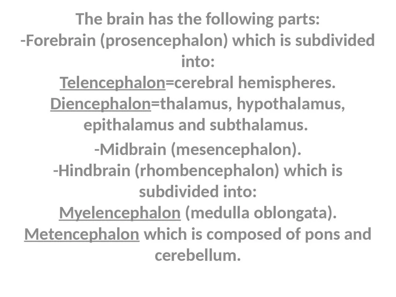
/
The brain has the following parts: The brain has the following parts:

The brain has the following parts: - PowerPoint Presentation

FuzzyWuzzyBear
FuzzyWuzzyBear . @FuzzyWuzzyBear
Follow
342 views
Uploaded On 2022-08-03

The brain has the following parts: - PPT Presentation

Forebrain prosencephalon which is subdivided into Telencephalon cerebral hemispheres Diencephalon thalamus hypothalamus epithalamus and subthalamus Midbrain mesencephalon ID: 933824

cerebral sulcus lobe lateral sulcus cerebral lateral lobe central brain hemisphere surface parts rami gyri occipital fissure situated composed

Share:

Link:

Embed:

Download Presentation from below link

Download Presentation The PPT/PDF document "The brain has the following parts:" is the property of its rightful owner. Permission is granted to download and print the materials on this web site for personal, non-commercial use only, and to display it on your personal computer provided you do not modify the materials and that you retain all copyright notices contained in the materials. By downloading content from our website, you accept the terms of this agreement.


Presentation Transcript

Slide1

The brain has the following parts:

-Forebrain

(

prosencephalon

) which is subdivided into:

Telencephalon

=cerebral hemispheres.

Diencephalon

=thalamus, hypothalamus,

epithalamus

and

subthalamus

.

-Midbrain

(mesencephalon).

-Hindbrain

(

rhombencephalon

) which is subdivided into:

Myelencephalon

(medulla oblongata).

Metencephalon

which is composed of pons and cerebellum.

Slide2

Slide3

General facts:

֍glial cells are ten times more than neurons in the mammalian brain.

֍there is no connective tissue in the CNS.

֍The mid brain, pons and medulla oblongata will form the brain stem.

֍There are no two identical brains.

֍Cortex = grey matter = gyri and sulci.

֍

medulla = White

matter ,

it contains group of nuclei (grey matter) inside it.

Slide4

Topography of the telencephalon:

There

are

right and left cerebral hemispheres, they form the largest part of the brain. They are incompletely separated by the longitudinal cerebral fissure that contains the

falx

cerebri

of the

dura

mater

each cerebral hemisphere possesses a central cavity called the lateral ventricle.

Slide5

Slide6

Slide7

Each cerebral hemisphere has three

surfaces:

-

Superolateral

surface

-medial surface

-inferior surface

Slide8

Slide9

Slide10

Slide11

Superolateral surface:

The central sulcus

: just behind the mid point of the superior border of the hemisphere, the fissure of Rolando (central sulcus) is start.

Slide12

The lateral sulcus

: (fissure of

Sylvius

) it is composed of anterior and posterior rami or parts. At the junction of the two parts, there are two ascending rami (V- shaped rami) arise toward the inferior frontal

gyrus

, and so this

gyrus

is further divided by this V- shaped ascending rami into pars

orbitalis

, pars

triangularis

and pars

opercularis

.

Slide13

Slide14

Insula (island of

Reil

)

: is situated deep in the floor of the lateral sulcus and is almost surrounded by a circular sulcus, and can only be seen when the lips of the lateral sulcus are widely separated. It involves parts from frontal, parietal and temporal lobes. The insula is composed of long and short gyri that almost completely surrounded by the circular sulcus. The insula is divided into two regions by a central sulcus, several short gyri lie in front of the central sulcus and one or two long gyri lie behind it.

Slide15

Slide16

Lobes of the cerebral hemisphere:

Frontal lobe: it is situated in front of the central sulcus and above lateral sulcus.

Parietal lobe: it lies behind sulcus and above the lateral sulcus.

Temporal lobe: it is below the lateral sulcus.

Occipital lobe: it is situated below and behind the

parieto

-occipital sulcus. Pre-occipital notch is the land mark that delineates the occipital lobe from the temporal lobe.

Slide17

Medial surface of the cerebral hemisphere has the main following features:

The corpus callosum:

the two cerebral hemispheres are joined by the corpus callosum which formed from rostrum anteriorly, genu, body and

splenium

posteriorly

.

The

calcarine

sulcus: in which the visual cortex is around this sulcus.

Slide18

Slide19