PPT-Quantitative CT Evaluation of Small Pulmonary Vessels Has Functional and Prognostic Value
Author : Heartbreaker | Published Date : 2022-08-03
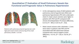
Shahin Y et al Published Online July 12 2022 httpsdoiorg101148radiol210482 In this retrospective study of 1823 patients with pulmonary arterial hypertension PAH
Presentation Embed Code
Download Presentation
Download Presentation The PPT/PDF document "Quantitative CT Evaluation of Small Pulm..." is the property of its rightful owner. Permission is granted to download and print the materials on this website for personal, non-commercial use only, and to display it on your personal computer provided you do not modify the materials and that you retain all copyright notices contained in the materials. By downloading content from our website, you accept the terms of this agreement.
Quantitative CT Evaluation of Small Pulmonary Vessels Has Functional and Prognostic Value: Transcript
Download Rules Of Document
"Quantitative CT Evaluation of Small Pulmonary Vessels Has Functional and Prognostic Value"The content belongs to its owner. You may download and print it for personal use, without modification, and keep all copyright notices. By downloading, you agree to these terms.
Related Documents