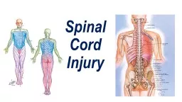
Dr Raj Kumar Yadav Assist Prof PMR MBBS 180719 Every slide has 4 to 5 statements Out of these 1 statement is false Identify it SCI is a devastating life threatening event Currently 225000228000 individuals living in US with sequelae of SCI including permanent paralysis ID: 930311
Download Presentation The PPT/PDF document "Acute Spinal C ord Injury" is the property of its rightful owner. Permission is granted to download and print the materials on this web site for personal, non-commercial use only, and to display it on your personal computer provided you do not modify the materials and that you retain all copyright notices contained in the materials. By downloading content from our website, you accept the terms of this agreement.
Slide1
Acute Spinal Cord Injury
Dr
Raj Kumar
Yadav
Assist. Prof., PMR
MBBS 18/07/19
Slide2Every slide has 4 to 5 statements.
Out of these 1 statement is false
Identify it
Slide3SCI is a devastating life threatening event.
Currently 2,25000-2,28000 individuals living in U.S. with sequelae of SCI including permanent paralysis.
Male: female- 1:4
Age: 16-30
yrs
majority
Slide4Leading causes-
motor
vehicle
accidents- 47.5
%
sports
-22.9%
violance-13.8
%
Falls- 8.9
%
Slide5Acute
SCI: complex, multifaceted
.
Mechanical trauma cause direct neuronal
damage
However
a
small
no. of axons are lost as a result of secondary pathophysiological
events-
hypo
perfusion, ischemia, and biochemical and inflammatory changes
Salvaging as little as 10% of adult axons can makes walking a potential goal.
Slide6Common Injuries
Slide7C-Spine Flexion Injury
Occurs during Cervical
flexion
with axial loading
C5- least commonly injured
Anterior
wedging
+/-
retropulsion
of
bony fragments into spinal canal is present
Slide8Cervical - Facet dislocations
Unilateral
Occurs in Flexion/rotation injury,
C5-C6 is
most common
More
likely to be
complete
• Bilateral
Occurs in Flexion injury,
C5-C6 is most common
More likely to be complete
Slide9Cervical hyperextension injury
Occurs in Acceleration-deceleration
injury
Due to Falls
,
MVCs
C4-C5
most
common involvement
Do not Often
results in a central cord syndrome
Slide10Jefferson Fracture
It is C1
burst
fracture
Usually UNSTABLE
with
no
neuro
findings
Due to Axial
loading of
atlasCommon in contact sports
Slide11Hangman Fracture
C1
burst
fracture
Bilateral
fracture
from deceleration injury
Common in Head
hitting
windshield
Mostly stable
Slide12Chance Fracture
It is T12-L2
transverse
fracture through
posterior
elements and
vertebral
body
Common in lap
belt
injury
Caused by Hyperextension of thorax4. Degree of injury depends on
movement
of bony elements
Slide13Management at Injury Site
Slide14Critical factors in recovery:
Late pre hospital recognition of injury
Prompt resuscitation
Stabilization of injury
Avoidance of additional neurological injury and medical complications.
Slide15Prehospital management
- 3
to 25 percent of SCIs occur after the
initial traumatic
insult, either during transit or early in the course of
treatment.
Four responsibilities of
prehospital
(infield) care are:
initial evaluation
a
dequate resuscitationmobilization of the suspected fractured area
s
afe extrication, and transportation
Slide16Initial Evaluation
Steps of Trauma PRIMARY SURVEY
are:
A
irway maintenance with cervical spine control;
B
reathing and ventilation management;
C
irculation with hemorrhage control;
D
isability (neurological status) limitation
E
xposure/
E
nvironmental control (covering the patient while preventing hyper- or hypothermia)
Slide17Evaluation
Secondary survey includes:
a detailed Head to toe evaluation
Quick motor examination : grip strength & a foot dorsiflexion evaluation
Gross sensory examination.
Signs of incontinence, urinary retention, priapism, or loss of anal sphincter tone are usually not found in SCI.
Slide18All
evaluations must take place in full spinal
immobilization
In an unconscious patient assume that cervical spine is injured until radiography of its entire length prove otherwise.
Even
in the absence of any of the
clinical
findings, the patient must be placed in a rigid collar and backboard and immobilize for transport.
Resuscitation begins
after
the
secondary
survey
Slide19IMMOBILIZATION
All major trauma
victims must be
immobilized
P
atients complaining of neck pain or neurological
symptoms must be
immobilized
A
ny patient with altered mental status of uncertain cause must be immobilized.
Flexed position of spine is critical to prevent any further damage to the cord.
Slide20Immobilization
Secure the neck first by a cervical collar.
When
removing a patient from a seated position, a cervical collar is first placed on the
patient.
Immobilize
the entire spine using a
soft
board.
Slide21Slide22Extrication and Transportation
After proper immobilization, a safest method of extrication and transportation should be adopted.
After proper immobilization, a
fastest
method of extrication and transportation should be
adopted.
Minimum Three persons are required during extrication
S
coop stretcher &
Kendric
extrication devices(KED) are used for extrication
Slide23Scoop stretcher & KED
Slide24Patient on vacuumed mattresses
Slide25Goals of Medical Management in a hospital:
1. Normalize vital signs.
2. Minimizing the neurological damage caused during the primary injury
3. Prevent aspiration
4. Preventing further cord injury secondary to hypo perfusion, ischemia, and biochemical and inflammatory changes
Slide26Management in Hospital
Slide27Spinal Stability- neurological and mechanical:
Neurologic stability denotes a state in which, under the stresses that are imposed, no further neural damage is caused.
Mechanical stability refers to the relative motion of vertebral segments under the physiologic loads of everyday activity.
Slide28Spinal Stability
To assess mechanical stability: Dennis’ 3 column theory used.
A column can be disrupted by either fracture or ligamentous disruption.
Disruption of three or more columns imparts instability.
Flexion & Extension X-ray films are done in conscious patients with no neurological deficits.
Slide29Imaging:
Computerized tomography (CT):
provides highly detailed axial images of each vertebral segment
inferior to MRI for delineating fractures.
can demonstrate
nondisplaced
fractures not discernible on plain x-ray.
an excellent method for evaluating and quantifying the degree of spinal canal compromise.
Slide30Imaging
MRI:
M
odality of choice for evaluating the spinal cord and neural elements.
C
an reveal edematous soft tissues, in either the anterior or posterior cervical spine.
Do not offer information regarding the integrity of the ligamentous structures
Can detect presence of herniated disc material in the spinal canal or foramina
Slide31Closed Reduction and Immobilization
Most mechanically stable thoracolumbar injuries without neurologic deficit can be treated
nonoperatively
Nonoperative
treatment should not be considered in stable injury patterns with limited potential for progressive deformity and neurologic compromise.
Slide32Operative Treatment
The indications for surgical intervention of spinal cord injuries depend on
pattern of injury.
alignment and stability of the vertebral fracture.
neurologic status of the patient.
overall medical condition of the patient do not affect decision
Slide33T
SURGICAL Management:
decompress the neural elements by anterior decompression or Posterior decompression
spine stabilized by instrumentation.
spinal
orthosis
- for 1 month
.
Slide34TREATMENT MODALITIES
METHYLPREDNISOLONE-
stabilize membranes, inhibit lipid peroxidation, suppress
vasogenic
edema by restoring the blood-central nervous system (CNS) barrier
enhance the spinal cord blood flow, inhibit pituitary endorphin release, and attenuate the inflammatory response.
Timing of steroid therapy is critical in its ultimate efficacy (8-72
hrs
)
recommended for penetrating
SCI.
Slide35SCI is followed by a series of detrimental hemodynamic and biochemical processes
that cannot be prevented by early and aggressive medical management
PREVENTION &TREATMENT OF COMPLICATIONS
Slide36Cardiovascular picture following SCI:
Spinal shock - loss or depression of all or most spinal reflex activity below the level of the injury
Hypotension-
common in lower level of injury, caused by a withdrawal of sympathetic tone.
Neurogenic shock - The vasodilatation, hypotension, decreased peripheral vascular resistance (PVR), decreased preload, and bradycardia.
Slide37Deep Venous Thrombosis: Prevention and Treatment
Causes:
Immobility
vascular dilatation and stasis
epithelial damage, and
an increase in the level of factor VIII and fibrinogen.
Slide38Incidence
of DVT during acute inpatient stay: 13.6
%.
pneumatic
devices
are applied
to the lower extremities for the first 2 weeks after the
injury.
If
thromboprophylaxis
is delayed for more than 72
hours, venous doppler to screen for thrombi formation prior to application of above devices are usually not required
Slide39Deep Venous Thrombosis: Prevention and Treatment
Anticoagulant prophylaxis with LMWH are initiated after 72 hours
It is continued until discharge in patients with incomplete injury.
for 8 weeks in patients with uncomplicated complete injury.
for 12 weeks or until discharge from rehabilitation in Complicated Complete injury.
Slide40AUTONOMIC DYSREFLEXIA
Occurs at Neurological level-T6 and above
Characterized by
a
brupt onset of malignant hypertension &
bradycardia
Caused by any noxious stimulus
Only treatment possible is to give
Nifedipine
& nitrates
Treatment is to Identify & treat noxious stimulus
Slide41Respiratory Management
Complete SCI with neurological level C4- ventilator dependent
C3 injuries- borderline
C2 and above- ventilator dependent
Primary Goal- Recruiting and maintaining aeration of alveoli thereby preventing atelectasis and pneumonia.
Slide42Atelectasis & pneumonia:
Occur 40-70% of
tetraplegics
Most commonly occurs in the first 5 to 7 days
Often focused primarily in the left upper lobe
Chest percussions, postural drainage, assistive coughing, Intermittent positive pressure breathing, Bronchodilators & mucolytic agents(
guaifenesin
) are used for treatment
Slide43Gastrointestinal Care
During spinal shock: gastric dilatation and paralytic ileus leads to distended stomach
vomiting and aspiration.
nasogastric tube is used for distended stomach
Stress ulcers are rare occurring in the acute phase following SCI
Prophylactic therapy with H2 blockers should be instituted for stress ulcers
Bowel management program should be established once normal bowel sounds and motility are restored.
Slide44Bladder Care
During spinal shock: bladder distension leads to urinary reflux which can result in renal failure
A Foley’s catheter should be inserted at admission.
During
subacute
phase of injury, use of Clean Intermittent Catheterization (CIC) increases the risk of bacterial infections.
Slide45The
most frequent secondary medical complication reported during the acute care of SCI patients is urinary tract
infection.
Symptomatic UTI should be treated with appropriate
antibiotics for 7 to 14 days.
Asymptomatic
bacteriuria
should
be treated routinely
Slide46Skin Care
Pressure ulcers are a
devastatating
complication of SCI (40%)
Posture change is required every 2 hourly.
Specially designed foam/air mattresses can reduce the pressure over bony prominences, but will not obviate the need for turning.
Adequate nutrition is not important for pressure ulcer healing
Enteral
rather than
parenteral
nutrition
is preferred once
patient stabilized
Slide47Ambulatory training
Slide48Tilt Table
Slide49Strengthening of UL & Trunk
Slide50Standing balance
Slide51Orthotic support
Slide52Robotic-assisted Gait Training (
Lokomat
)
Slide53Robotic-assisted Upper Limb Training (ARMEO)
Slide54Virtual Reality-based Training (Nirvana)
Slide55THANKS