/
Back  muscles ,  occipital Back  muscles ,  occipital

Back muscles , occipital - PowerPoint Presentation

HoneyBun
HoneyBun . @HoneyBun
Follow
343 views
Uploaded On 2022-08-02

Back muscles , occipital - PPT Presentation

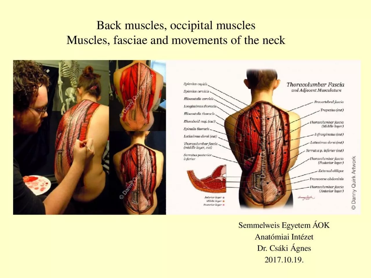
muscles Muscles fasciae and movements of the neck Semmelweis Egyetem ÁOK Anatómiai Intézet Dr Csáki Ágnes 20171019 The muscles of the back can be divided into three groups ID: 932362

processes muscles posterior layer muscles processes layer posterior movements transverse spinous vertebral ribs neck capitis innervation fibres spinal part

Share:

Link:

Embed:

Download Presentation from below link

Download Presentation The PPT/PDF document "Back muscles , occipital" is the property of its rightful owner. Permission is granted to download and print the materials on this web site for personal, non-commercial use only, and to display it on your personal computer provided you do not modify the materials and that you retain all copyright notices contained in the materials. By downloading content from our website, you accept the terms of this agreement.


Presentation Transcript

Slide1

Back muscles, occipital musclesMuscles, fasciae and movements of the neck

Semmelweis Egyetem ÁOKAnatómiai IntézetDr. Csáki Ágnes2017.10.19.

Slide2

The muscles of the back can be divided into three groups :Superficial – associated with movements of the shoulder. Spinohumeral muscles M. trapezius M. rhomboideus major et minor

M. levator scapulae

M.

latissimus dorsiIntermediate – associated with movements of the thoracic cage. Serratus posterior superior Serratus posterior inferiorDeep – associated with movements of the vertebral column erector spinae m. transversospinal muscles, interspinal, intertransverse and suboccipital musclesThe deep muscles develop embryologically in the back, and are thus described as intrinsic muscles. The superficial and intermediate muscles do not develop in the back, and are classified as extrinsic muscles

Back

muscles

Slide3

differentiation of the myotome – development of the skeletal musclesEpimere

– muscles of the back(segmental innervations from the dorsal branch of the spinal nerve)Hypomere – muscles of the neck, thorax and abdomen, muscles of the extremities

(innervated by ventral branches of the spinal nerve - plexus)

Slide4

A very similar layered organization at the thorax and abdomen

Slide5

M. erector spinae----------

---------- M. rectus abdominis------------ Mm obl. abd. ext. et int.

--------------------

Mm. scaleni

Slide6

The muscles of the back can be divided into three groups :Superficial – associated with movements of the shoulder. Spinohumeral muscles migrate from the hypomere M. trapezius M. rhomboideus

major et minor M. levator scapulae M. latissimus

dorsi

Intermediate – associated with movements of the thoracic cage. Serratus posterior superior Serratus posterior inferiorDeep – associated with movements of the vertebral column epimere erector spinae m. transversospinal muscles, interspinal, intertransverse and suboccipital musclesBack muscles

Slide7

Superficial muscles

– associated with movements of the shoulder.Trapezius muscleOriginates from the skull(nuchal

line)

, and the spinous processes of C1-T12. The fibres attach to the clavicle, acromion and the scapula spine.Innervation : XI. cranial nerve (accessory nerve)

Proc. spinosus

M. trapezius

M.

latissimus

dorsi

Vertebra prominens, proc. spinosus

The upper

fibres

of the

trapezius

elevates the scapula and rotates it during abduction of the arm. The middle

fibres

retract the scapula and the lower

fibres

pull the scapula inferiorly.

Slide8

P

roc. spinosus

M.

latissimus dorsiVertebra prominens, proc. spinosusLatissimus dorsibroad origin from the spinous processes of T6-T12, iliac crest, thoracolumbar fascia and the inferior three ribs attaches to the crest of lesser tuberculeInnervation: Thoracodorsal nerve (brachial plexus)Actions: Extends, adducts and medially rotates the upper limb.

Slide9

M.

rhomboideus

major

M. levator scapulaeM. rhomboideus minorLevator scapulae

from the transverse processes of the C1-C4 vertebrae and attaches to the

angle

of the scapula.

Innervation

:

Dorsal scapular nerve

(

brachial

plexus

)

Elevates the scapula

Rhomboid Major

from the

spinous

processes of T2-T5 vertebrae

,

a

ttaches

to the medial border of the scapula, between

the spine and

inferior angleRhomboid Minorfrom the spinous processes of C7-T1 vertebrae, attaches to the medial border of the scapula, Innervation: Dorsal scapular nerve (brachial plexus)Retract and rotate the scapula

Slide10

Serratus

posterior

superior m.Serratus posterior inferior m.Intermediate – associated with movements of the thoracic cage.

The

serratus

posterior superior is a thin, rectangular shaped muscle. It lies deep to the rhomboid muscles on the upper back.

Attachments

: Originates from the lower part of the

ligamentum

nuchae

, and the cervical and thoracic spines (usually C7 – T3). The

fibres

pass in an

inferolateral

direction, attaching to ribs 2-5.

Innervation

:

Intercostal

nerves

Actions

: Elevates ribs 2-5

The

serratus

posterior inferior is broad and strong. It lies underneath the

latissimus

dorsi.Attachments: Originates from the thoracic and lumbar spines (usually T11 – L3). The fibres pass in a superolateral direction, attaching to ribs 9-12.Innervation: Intercostal nervesActions: Depresses ribs 9-12.

Slide11

M.

splenius

capitisDeep – associated with movements of the vertebral columnM. splenius cervicisSplenius capitis Originates from the ligamentum nuchae, and the spinous processes of C7 – T3vertebrae. The fibres ascend, attaching to the the occipital bone of the skull.

Actions

: Rotate head to the same side.

Splenius

cervicis

Originates from the

spinous

processes of T3-T6

vertebae

. The

fibres

ascend, attaching to the transverse processes of C1-4.

Actions

: Rotate head to the same side.

Innervation

: Posterior rami of the spinal

nerves

The two splenius muscles can also act together to extend the head and neck.

Slide12

M.

iliocostalis

M

.

erector

spinae

:

The erector

spinae

is situated

posterolaterally

to spinal column, between the vertebral

spinous

processes and the costal angle of the ribs

.

It

has

three

columns

m.

iliocostalis

m.

longissimus

m.

spinalis

The

iliocostalis

muscle

is located laterally within the erector

spinae

. It is associated with the ribs, and can be divided into three parts –

lumborum

,

thoracis

, and

cervicis

.

Attachments

: Arises from the common

tendinous

origin, and attaches to the costal angle of the ribs and the cervical transverse processes.

Innervation

: Posterior

rami

of the spinal nerves

Actions

: Acts unilaterally to laterally flex the vertebral column.

Slide13

M.

longissimus

The longissimus muscle is situated between the iliocostalis and spinalis. It is the largest of the three columns. It can be divided into three parts – thoracic, cervicis and capitis.Attachments: Arises from the common tendinous origin, and attaches to the lower ribs, the transverse processes of C2 – T12, and the mastoid process of the skull.Innervation: Posterior rami of the spinal nervesActions: Acts unilaterally to laterally flex the vertebral column

Slide14

M. spinalis

The

spinalis

muscle is located medially within the erector spinae. It is the smallest of the three muscle columns. It can be divided into the thoracic, cervicisAttachments: Arises from the spinous processes and attaches to the spinous processesInnervation: Posterior rami of the spinal nervesActions: Acts unilaterally to laterally flex the vertebral column.

Slide15

M.

Iliocostalis

Lumborum

, thoracis, cervicisM. LongissimusThoracis,

cervicis

,

capitis

M.

Spinalis

Lumborum

,

thoracis

,

cervicis

M. longissimus capitis

Semispinalis

capitis

Slide16

M.

Semispinalis

cervicisTransversospinalis muscles are located underneath the erector spinae. They are a group of short muscles, associated with the transverse and spinous processes of the vertebral columnThere are three types of muscles in this group – the semispinalis, multifidus and rotatores.Semispinalis m.Originates from the transverse processes of C4-T10. The fibres ascend 5-6 vertebral segments

, attaching to the

spinous

processes of C2-T4

:

semispinalis

cervicis

and to the occipital bone of the skull

:

semispinalis

capitis

M.

Semispinalis

capitis

Slide17

Mm.

rotatores

Mm.

multifidiMultifidus m.Originates from the transverse processes The fibres ascend 2-4 vertebral segments, attaching the spinous processes of the vertebrae.Rotatores m.Originates from the vertebral transverse processes. The fibres ascend, and attach to the lamina and

spinous

processes of

the immediately superior vertebrae

.

Slide18

Minor Deep

Intrinsic Muscles:Levatores costarum –

Originates

from the transverse processes of C7-T11, and attaches to the rib immediately below. Acts to elevate the ribs.Interspinales: Spans between adjacent spinous processes. Acts to stablise the vertebral column.Intertranversarii– Spans between adjacent transverse processes. Acts to stablise the vertebral column.

Slide19

Erector spinaeOnly the semispinalis is shown

Slide20

Slide21

Suboccipital

muscles-

Muscular layers on the neck

Slide22

M.

rectus

capitis post. Minorfrom the posterior tubercle of the C1 vertebra (atlas) to the medial part of the inferior nuchal line of the occipital bone. Obliquus capitis superior

rom

the transverse process of C1 and attaches into the occipital bone (between the superior and inferior

nuchal

lines).

M.

obliquus

capitis

inferior

from the posterior tubercle of the C2 vertebra, and attaches into the transverse process of C1.

M.

rectus

capitis

post.

Major

from the

spinous

process of the C2 vertebrae (axis), and inserts into the lateral part of the inferior

nuchal

line of the occipital bone.

Suboccipital

muscles-

suboccipital

trigone

Actions

: Extension and rotation of the head.

Innervation

Suboccipital

nerve (post

.

ramus

of C1)

Slide23

Prevertebral muscles of the neck

M.

longus

colliM. longus capitisM. rectus capitis anteriorM. rectus capitis lateralis

Actions

:

flex

the

head

and

neck

anteriorly

Innervation

ventral

rami

of

spinal

nerves

Scalenus

muscles

Slide24

NameOrigin

Insertion

Function

m. anterior scalene scalene tubercle on the 1. ribanterior tubercles of the transverse processes of C3-C6

The

three

muscles

:

ipsilateral

contraction

causes

ipsilateral

lateral

flexion

of

the

neck

,

and

bilateral

contraction

causes anterior

flexion

of

the

neck

in

case

of

forced

respiration

act

as

accessory

muscles

of

respiration

innervation

:

anterior

rami

of

spinal

nerves

C3-C8

m. middle scalene

behind

the subclavian groove on the 1. rib

posterior tubercles of the transverse processes of C2-C7

m. posterior scalene

2. rib

posterior tubercles of the transverse processes of C5-C7

anterior

posterior

Scalenus

muscles

Slide25

Supra- and infrahyoid muscles The suprahyoid muscles are a group of four muscles, located superiorly to the hyoid bone of the neck. They all act to elevate the hyoid bone – an action involved in swallowingThe

infrahyoid muscles are a group of four muscles that are located inferiorly to the hyoid bone in the neckDepress the

hyoid

bone.Innervation: ventral rami of spinal nerves (ansa cervicalis)

Slide26

Lateral view of supra- and infrahyoid muscles

Slide27

Superficial neck musclesSternocleidomastoid m.Originates from the manubrium sterni and

clavicle, inserts to the mastoid process of the

skull

ipsilateral contraction causes ipsilateral lateral flexion and rotation of the head („ear comes near to the shoulder”bilateral contraction causes protraction of the headInnervation: IX cranial nerve (with the

trapezius

m.)

Slide28

Cervical fasciaFascia is a layer of fibrous tissue that surrounds muscles, vessels and nerves. In the neck, there are several layers of fascia, which act to support and compartmentalise the structures presentSuperficial layerPretracheal layer

Prevertebral layer

Slide29

Fascia

cervicalis

- Spf. Layer or investing layer- pretracheal layer (visceral part) (carotid sheath)- prevertebral layerThe investing layer of fascia in highlighted in blue. T

he fascia completely envelopes the

sternocleidomastoid

and

trapezius

.

Transverse section of the neck.

Slide30

Fascia

cervicalis

- Spf. Layer or investing layerpretracheal layer(muscular) (visceral) (carotid sheath)- prevertebral layer

The trachea,

esophagus

, and

infrahyoid

muscles

are

enclosed

by

the

pretracheal

layer

.

It

can

be

anatomically divided into two parts:Muscular – encloses the infrahyoid muscles(purple)Visceral  – encloses the

trachea and oesophagus and forms the carotid sheath

Slide31

Fascia

cervicalis

- Spf. Layer or investing layer- pretracheal layer (visceral part) (carotid sheath)- prevertebral layer

The

prevertebral

layer

surrounds the 

vertebral

column

and its associated muscles (scalene,

prevertebral

, and deep muscles of the back)

Slide32

The superficial layer and muscular part of pretracheal layer end at the sternum, but the visceral layer and the

prevertebral layer continous downward to the thorax!! -

inflammation

Slide33

Sternocleidomastoideus (elevates clavicle)Nasal muscles (

nostrils widen)

Scalenus anterior, medius et posterior

(elevates upper ribs)Auxiliary respiratory muscles

Slide34

Serratus post. sup. (elevates the upper ribs)

Levator costarum (elevates the ribs)

Auxiliary

respiratory musclesSerratus post. inf. (depresses the lower ribs)Quadratus lumborum (depresses the XIIth rib)

Slide35

Movements of the cervical part

Articulatio atlantooccipitalis articulatio

ellipsoidea

movements: flexio lateralis flexio ventralis és dorsalis Articulatio atlantoaxialis articulatio trochoidea movements: ¾ of rotation of the neck!!!Cervical spine direction of the articular surfaces: horizontal movements: flexio lateralis + small rotation flexio ventralis and dorsalis

Slide36

Thoracic part direction of the articular surfaces: oblique movements:

small torsion flexio lateralis Lumbal part direction of the articular surfaces: sagittal

movements:

flexio ventralis and dorsalis flexio lateralis Movements of thoracic and lumbal part

Slide37

DorsalflexionMovements of the spineClordosis

Tkyphosis

L lor-

dosisC.-L. lordosis decreasedTh. kyphosis increasedC.-L. lordosis increasedTh. kyphosis decreasedvery: between the C.-Th. and L.-S. borderlandsvulnerable !AnteflexionLateralflexionC.-L.Th. hardlyRotation:-C. noLower Th. -L. hardly

Slide38

Irodalom:images.MD, 2006 Current Medicine LLC Kahle W, Leonhardt H, Platzer W: Color Atlas/Text of Human Anatomy

, 1992, Thieme, Stuttgart Putz R, Pabst

R

(editors): Sobotta Atlas of Human Anatomy, 1993, Urban & Schwarzenberg, MünchenRomanes GJ (editor): Cunningham’s Textbook of Anatomy, 1991, Oxford University Press, OxfordSzentágothai J, Réthelyi M: Funkcionális anatómia, 2002, Medicina, BudapestVízkelety T: Az ortopédia tankönyve, 1995, Semmelweis Kiadó, Budapesthttp://teachmeanatomy.info/the-basics/https://www.youtube.com/watch?v=F2sSVWEdjWQThank you!