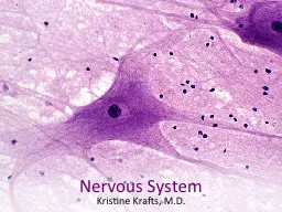
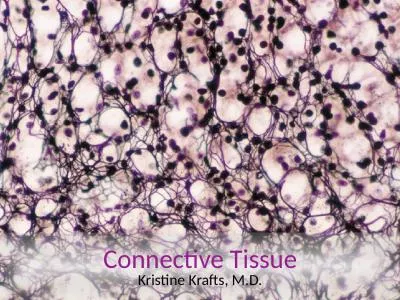
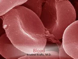
The most beautiful thing we can experience is the mysterious It is the source of all true art and science Albert Einstein Blood Lecture Objectives Be able to identify and describe the major functions of the following ID: 935221
Download Presentation The PPT/PDF document "Blood Kristine Krafts, M.D." is the property of its rightful owner. Permission is granted to download and print the materials on this web site for personal, non-commercial use only, and to display it on your personal computer provided you do not modify the materials and that you retain all copyright notices contained in the materials. By downloading content from our website, you accept the terms of this agreement.
Slide1
Blood
Kristine Krafts, M.D.
Slide2The most beautiful thing we can experience is the mysterious.
It is the source of all true art and science.
-Albert Einstein
Slide3Slide4Slide5Slide6Blood Lecture ObjectivesBe able to identify and describe the major function(s) of the following: Erythrocytes (RBCs
)Granulocytes (neutrophils, eosinophils, basophils)Agranulocytes (lymphocytes, monocytes) Platelets Know the approximate percentage
of each type of leukocyte present in normal blood.Be able to describe the differences between plasma and serum.
Slide7Blood Lecture OutlineIntroduction Erythrocytes
PlateletsLeukocytesGranulocytesAgranulocytes
Slide8Blood Lecture OutlineIntroduction
Slide9Blood is a Specialized Connective TissueComposed of:CellsPlasma
Slide10CellsRed cells (erythrocytes)White cells (leukocytes)
Platelets
Slide11Plasma contentsWater: 92%Proteins: 7% Albumin: 58%
Immunoglobulins: 37%Fibrinogen: 4%Other proteins: 1%Other stuff: 1% (electrolytes, nutrients, respiratory gases, waste products)
Slide12Plasma vs. SerumPlasma clots, serum does not clotSerum = plasma minus clotting factors (it’s what’s left after plasma clots)
Slide13Blood Lecture OutlineIntroduction
Erythrocytes
Slide14Erythrocytes (Red Blood Cells)Life span: 120 daysDerived from red cell precursors in bone marrowNormal numbers
Male: 4.5-6 x 10¹²/LFemale: 4-5 x 10¹²/L
Slide15Normal
red blood cells
Slide16Red Blood Cell Morphological FeaturesNicely designed biconcave disk shape
Roughly 7 µm wide and 2 µm thickCytoskeleton: spectrin, ankyrin, actinNo nucleusCytoplasm: water (65%); organelles (1%); hemoglobin (34%)
Slide17Slide18⅓
Pliable membrane allows cells to squeeze through tiny spaces.
Slide20z
Slide21Main red cell function: transport oxygen using hemoglobin
Hemoglobin
4 globin chains
4 heme molecules
Heme molecule
(carries O
2
)
Slide22Blood Lecture OutlineIntroduction
ErythrocytesPlatelets
Slide23PlateletsLife span: 8-10 daysDerived from megakaryocytes in bone marrow
Normal number: 150-450 x 109/LAbout 2 µm in diameterGranulomere and hyalomere regionsNo nucleus
Function: help blood to clot
Slide24Normal platelets
Slide25Normal Platelets
Slide26Platelets look boring but have a ton of stuff inside (granules) and outside (receptors)
Slide27Platelets forming a clot
Slide28Blood Lecture OutlineIntroduction
ErythrocytesPlateletsLeukocytes
Slide29White blood
cells (nice drawing)
Slide30White blood
cells (real blood smear)
Slide31White blood
cells (another real blood smear)
Slide32White blood cell count (WBC)Just gives you the total number of white blood cells (normal is about 4-11 x 10
9/L).
Slide33White blood cell differential (“diff”)
Tells you how many of each type of white cell are present (normally, neutrophils are the most numerous, and basophils are the least numerous).
Slide34LeukocytesGranulocytesNeutrophilsEosinophils
BasophilsAgranulocytesLymphocytesMonocytes
Slide35Wait, agranulocytes have granules?!Yes! Both granulocytes and agranulocytes have cytoplasmic granules called azurophilic granules.
But granulocytes also have specific granules that define them as cells (neutrophilic, eosinophilic and basophilic granules)
Slide36Granulocytes
vs.
agranulocytes
Slide37Blood Lecture OutlineIntroduction
ErythrocytesPlateletsLeukocytesGranulocytes
Slide38Neutrophils45-75% of differential count (between 2-8 x 10⁹/L)
About 15 µm in diameterMulti-lobed nucleus… …hence their other name: “polymorphonuclear leukocyte” (PMN)Two kinds of granules:
Azurophilic (primary, purple) granules (a few)Neutrophilic (secondary, pink) granules (lots)
Slide39Normal neutrophils
Immature neutrophil
Slide40Neutrophil: azurophilic vs. neutrophilic
granules
(hard to see on screen!)
Slide41Normal neutrophil
(top left)
Neutrophil
from patient with bacterial infection
Azurophilic granules become much more
prominent during bacterial infection.
Slide42Azurophilic granules first appear in less mature neutrophils called promyelocytes. Promyelocytes divide, distributing their azurophilic granules evenly (which
means more mature
neutrophils have fewer azurophilic granules).
promyelocyte
Slide43Neutrophil FunctionsFirst line of defense against invaders (bacteria, foreign objects)Spend a few hours in blood, then migrate quickly to site of infection where they spend a few days
Kill invaders by phagocytosis and by enzymatic destruction (nasty!)Then take off and let others (macrophages) clean up the mess
Slide44Eosinophils1-4% of differential count (about 0.5 x 10⁹/L)About 15 µm in diameter
Large, gorgeous, orange-red (eosinophilic) granules in cytoplasmGreek eos = first blush of dawnBi-lobed nucleus
Slide45Eosinophil
Slide46Eosinophil in real life
Slide47Eosinophil FunctionsMajor cell involved in allergic reactions (like hay fever and asthma)Good at killing parasites (granules contain major basic protein)
Also involved in drug reactionsHelp modulate immune responses
Slide48BasophilsLess than 1% of differential count (less than
0.3 x 10⁹/L) About 10 µm in diameterTons of large, deep blue (basophilic) granules in cytoplasmIrregularly-shaped nucleus (hard to see under all those granules
)Functions: fight infection, mediate allergic responses
Slide49Basophil
Slide50Basophil in real life
Slide51Blood Lecture OutlineIntroduction
ErythrocytesPlateletsLeukocytes
GranulocytesAgranulocytes
Slide52Lymphocytes20-50% of differential count (between 1-4 x 10⁹/L)
Most lymphocytes are small (6-12 µm) but some are larger (up to 20 µm)Nucleus: dark staining; “clumpy and smudgy”Two main types (which look pretty much the same):
B-lymphocytesT-lymphocytes
Slide53Normal lymphocytes
Slide54Small lymphocyte in real life
Slide55Lymphocyte chromatin pattern: clumpy
and
smudgy
Slide56Neutrophil chromatin
: distinct clumps (with white space between clumps)
Lymphocyte chromatin
: clumpy but also smudgy (no white space between clumps)
Monocyte chromatin
:
not really clumpy
Slide57B-Lymphocytes15% of circulating lymphocytesDevelop in b
ursa of Fabricius (in birds) and in bone marrow (in humans)Further maturation occurs in lymphatic tissues (lymph nodes and spleen)Ultimately, become either plasma cells (which make antibodies) or memory cells (which “remember” previous infections)
Slide58T-LymphocytesAbout 85% of circulating lymphocytesDevelop and mature in t
hymus Also found in bone marrow and lymphoid tissues, along with B cellsUltimately, most become either cytotoxic T cells (which kill infected cells) or helper T cells (which help other immune cells do their jobs)
Slide59Monocytes1-8% of differential count (between 0.1-0.8 x 10⁹/L)
12-20 µm in diameterNucleus: indented, oval, kidney, or horseshoe-shaped. “Raked” chromatin.Cytoplasm: “dishwater” (gray-blue) color, sometimes with little vacuoles and/or tiny azurophilic granules
Slide60Monocyte: large cell with “dishwater” cytoplasm
and “raked” chromatin
Slide61Monocyte FunctionDifferentiate into macrophages (histiocytes) in different organsForeign body giant cells (anywhere)
Kupffer cells (liver)Microglial cells (brain)Alveolar macrophages (lung)Second line of defense against invading organismsHelp lymphocytes do their job; also phagocytic (eat up invaders and either get rid of them or present bits of them to lymphocytes)
Slide62Blood Lecture OutlineIntroduction Erythrocytes
PlateletsLeukocytesGranulocytesAgranulocytes