/
In the name of God Mucormycosis In the name of God Mucormycosis

In the name of God Mucormycosis - PowerPoint Presentation

NoPainNoGain
NoPainNoGain . @NoPainNoGain
Follow
342 views
Uploaded On 2022-08-01

In the name of God Mucormycosis - PPT Presentation

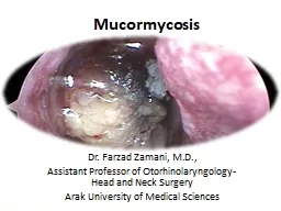
Drhmazaherpour Assistant professor of infectious disease AUMS A 38yearold male was admitted to our Hospital a tertiary care center with a history of fever for 4 days He presented with a high grade fever body ache cough and shortness ID: 931875

treatment mucormycosis therapy patients mucormycosis treatment patients therapy day posaconazole amb infection dose antifungal sinus formulation amphotericin days disease

Share:

Link:

Embed:

Download Presentation from below link

Download Presentation The PPT/PDF document "In the name of God Mucormycosis" is the property of its rightful owner. Permission is granted to download and print the materials on this web site for personal, non-commercial use only, and to display it on your personal computer provided you do not modify the materials and that you retain all copyright notices contained in the materials. By downloading content from our website, you accept the terms of this agreement.


Presentation Transcript

Slide1

In the name of God

Mucormycosis

Dr.h.mazaherpour

Assistant professor of infectious disease AUMS

Slide2

A 38-year-old male was admitted to our Hospital, a tertiary care

center with a history of fever for 4

days. He presented

with a high grade fever, body ache, cough and shortness

of breath

. Nasopharyngeal swab was sent for RT-PCR which came

positive and

a diagnosis of COVID-19 was confirmed.

The patient had no

history of

diabetes or any other debilitating conditions and no relevant

family history

.

Slide3

On admission, the

deranged

investigations were: Neutrophil

count 83.1% (35–66%), Lymphocyte count 9.5% (24–44%), Fasting

blood sugar (FBS) 98 mg/

dL

(70-110 mg/

dL

), Post-prandial blood

sugar (PPBS

) 146 mg/

dL

(110-140 mg/

dL

), HbA1c 6.3% (

<

6%), Serum

Interleukin-6 37.93

pg

/mL (

<

6.4

pg

/mL), CRP 17.84 mg/L (0–6 mg/L),

D-Dimer 460 ng/mL (0–500 ng/mL).

Slide4

He was monitored in the Intensive Care Unit for 5 days and was

started on Inj.Remdesivir IV with a loading dose of 200 mg, followed

by 100

mg daily for 11 days. Methylprednisolone was given by IV

infusion, 80

mg/day in 240 mL saline at 10 mL/h for 18 days. Also

Inj.Dexamethasone

4 mg twice daily was given for 12 days as a part of

COVID-19 management

. Post-treatment FBS 125 mg/

dL

, PPBS 352 mg/

dL

and HbA1c

12.3%

Slide5

After 18 days, the patient complaint of swelling and pain in the left

eye. He was referred to the Department of Head and Neck Oncology

for the

same. On clinically examination, there was malaise, proptosis,

chemosis

, periorbital cellulitis and restricted medial gaze. Visual

acuity was

6/6 with partial

opthalmoplegia

and no nasal discharge was seen

Slide6

Slide7

MRI brain & orbit was done showing an ill-defined

heterogenous

soft

tissue signal intensity (

hypointense

on T1W-imaging),

polyploidal

mucosal thickening involving left maxillary and ethmoid sinuses was

seen. There was displacement of

adjacent medial

and inferior rectus.

Retrobulbar

soft tissue fat stranding

and edema

with resultant displacement of left eye ball anteriorly leading

to proptosis

was seen. It was also found to be closely abutting the left

optic nerve

Slide8

Slide9

Crusting

was noted over posterior aspect of inferior turbinate,

septum and

conchae. The sinuses were debrided and the specimen obtained

was sent

for culture sensitivity and histopathology.

On histopathology

examination,

aseptate

broad based hyphae and gram positive bacilli were seen and

hence Mucormycosis suspected.

The medical regime changed to

Inj. Amphotericin

B 300 mg/day,

eyedrops

Tobramycin BD

Slide10

Slide11

Slide12

Slide13

mucormycosis

Agents of mucormycosis are ubiquitous fungi in the environment that

are commonly found in decaying organic substrates, including bread,

fruits, vegetable matter, soil, compost piles, and animal excreta. These

fungi characteristically produce large, ribbon-like hyphae that are

irregular in diameter with only occasional

septae

, hence the characterization of these organisms as

aseptate

fungi.

Slide14

Spores ranging from 3 to 11 µm in diameter

are easily

aerosolized and dispersed and cause infections in humans

when inhaled

or introduced through a cutaneous or percutaneous route.

Acquisition of Infection

The primary mode of acquisition of mucormycosis is inhalation of

spores from environmental

sources.

Trauma, penetrating wounds,

burns, and direct injection of

sporangiospores

can cause infection

through a

cutaneous or percutaneous route.

Slide15

Patient Populations at Risk

The populations most commonly at risk for mucormycosis

include patients

with poorly controlled diabetes mellitus, prolonged

neutropenia, high-dose

corticosteroid

treatment,

or immunosuppressive

therapy associated

with transplantation, and/or elevated levels of free iron,

which enhances

fungal growth

Patients who develop mucormycosis in the absence of underlying

disease or immunosuppression at the time of infection frequently

have histories

of penetrating trauma, burns, surgery, or illicit IV drug

use before

the infection.

Slide16

Rhinocerebral

Infections

Rhinosinusitis, rhino-orbital, and

rhinocerebral

infections are classic

manifestations of human mucormycosis. Infection is initially localized

to the nasal

turbinates

and paranasal sinuses after inhalation of

spores but

can rapidly progress to the orbit (

sino

-orbital) or brain (

rhinocerebral

), particularly in patients with diabetic ketoacidosis or

profound neutropenia

.

Slide17

Clinical Presentation

Initial symptoms of sinus invasion by mucormycosis are

indistinguishable from

other more common causes of sinusitis. Sinus pain,

congestion, headache

, mouth or facial pain,

otologic

symptoms, and

hyposmia

or anosmia

are common

Involved tissues become red, then violaceous,

and finally

black with thrombosis and tissue necrosis. Necrotic eschars

of the

nasal cavity and

turbinates

, facial lesions around the nose,

and

exophytic

or necrotic lesions of the hard palate extending from

the maxillary

sinus are signs of rapidly progressing infection.

Slide18

Extension of sinus disease is primarily into contiguous structures.

Maxillary sinus infection extends into the hard palate, nasal cavity,

and ethmoid

sinus. Sphenoid disease invades the cavernous sinus,

contiguous temporal

lobe, and internal carotid artery in the siphon. Septic

emboli from

the carotid artery into the frontal and parietal lobes can occur.

Ethmoid sinus disease may invade the face or frontal lobe but easily

crosses the lamina

papyracea

into the orbit.

Slide19

Periorbital edema, ptosis, proptosis,

chemosis

,

and

preseptal

and orbital edema are early signs of orbital extension.

Pain and

blurring or loss of vision often indicate invasion of the globe

or optic

nerve.

Patients with extensive

rhino-orbital or

rhinocerebral

disease may present with trigeminal and ocular

motor nerve

palsy after cavernous sinus invasion

A bloody

nasal discharge

may be an early sign of nasal mucosal invasion.

Slide20

Radiology

Radiographic imaging is often suggestive of severe sinusitis but lacks

the specificity to diagnose

rhinocerebral

mucormycosis. Computed tomography (CT) of the

sinuses typically

reveals mucosal thickening, air-fluid levels, and bony erosion

Extraorbital

muscle thickening is often the first sign on CT or MRI of

orbital involvement and should prompt empirical antifungal therapy

until surgical exploration or biopsy of the sinus and orbits can be performed, which should be done as soon as possible.

Slide21

TREATMENT

Successful treatment of mucormycosis relies on timely diagnosis,

reversal of

underlying predisposing factors, early surgical

débridement

of

infected tissue

, and rapid initiation of effective high-dose systemic

antifungal therapy

.

Early diagnosis is critical to the outcome of mucormycosis

because small focal lesions can be surgically resected before the

lesions progress

to involve critical structures or distal organs

Slide22

Patients with suspected

rhinocerebral

mucormycosis should undergo

a thorough examination and “staging” of their disease, including CT

of the paranasal sinuses and lungs as well as endoscopic examination

of nasal

turbinates

with biopsy of any suspicious lesions or necrotic

eschars.

Slide23

Rapid correction

of predisposing

conditions, such as control of hyperglycemia, reversal

of ketoacidosis

, and rapid tapering of glucocorticoid therapy, are

critical for

reversing conditions that favor fungal virulence and dissemination.

Slide24

Antifungal Therapy

Historically, the drug of choice for the treatment of mucormycosis

was conventional amphotericin B

deoxycholate

(ABD), administered at the maximal tolerated doses of 1 to 1.5 mg/kg/day

.

Unfortunately, high

doses of conventional amphotericin B are usually not tolerated

for more

than several days before renal function deteriorates, especially

in patients

with diabetes or receiving concomitant nephrotoxic therapies.

Slide25

Lipid Amphotericin B Formulations

Lipid formulations of amphotericin B are safer than ABD for

long-term administration

and, in our opinion, are the preferred first-line

treatment for

severe mucormycosis.

In one of the few published case series, 24 patients with

mucormycosis

and

diabetes as the predominant underlying risk factors were

treated with

ABLC after failure or intolerance of the conventional ABD formulation

Slide26

The overall response rate (improvement or cure of infection)

was 71

% ,

with few reported toxic effects, even in

patients with

preexisting renal dysfunction.

Several case series have

reported the

successful treatment of mucormycosis with the liposomal

formulation of

amphotericin B, sometimes administered at high doses (i.e., 10

mg/kg/day

) for prolonged treatment courses

Slide27

The optimal dosing approach for L-AMB in patients with mucormycosis is frequently debated.

Preclinical pharmacokinetics/ pharmacodynamics

studies

in murine models of pulmonary mucormycosis that

simulated human

dosing have suggested that ABLC at 5 mg/kg/day or

L-AMB at

10 mg/kg/day results in rapid antifungal accumulation in the

lung, reduced

fungal burden, and improved survival.

Slide28

In one of the few randomized trials that compared standard doses

of L-AMB (3 mg/kg/day) to a higher dose-regimen (10 mg/kg/day for

14 days, then 3 mg/kg/day) for invasive aspergillosis, patients

randomized to

the higher-dosed L-AMB regimen failed to achieve higher

response rates

but experienced significantly higher rates of nephrotoxicity

and severe

hypokalemia

Slide29

The feasibility of high-dose (10 mg/kg/day) L-AMB treatment for

the initial treatment of mucormycosis was explored in a

multicentric

prospective French study of 40 patients with invasive mucormycosis

.

The planned treatment of 10 mg/kg/day was administered as an

infusion of

at least 2 hours for 4 weeks.

Slide30

Of importance, although 4 weeks of high-dose L-AMB was planned,

the average treatment duration at this dose was 13.5 days (range, 0–28

), with

40% of patients experiencing a doubling in the baseline

serum creatinine

and severe hypokalemia (serum potassium <3

mmol

/L).

Therefore considerations for using doses of L-AMB higher than 5 mg/

kg/day should take into account that a sizable proportion of patients

will develop renal injury requiring dosage reduction or possibly a

switch to

triazole

therapy after the first 1 to 2 weeks of therapy.

Slide31

Triazoles

The prospects for using

posaconazole

in the treatment of

mucormycosis

has

improved with the introduction of an extended-release tablet formulation with improved bioavailability compared with the older

suspension, and

an IV formulation solubilized in

sulfobutyl

ether β-

cyclodextrin

.

In an open-label

study evaluating

posaconazole

as salvage therapy (not initial therapy),

the overall

success rate of

posaconazole

(800 mg/day) was 70% in 24

patients, and

it was well tolerated with only minimal GI side effects.

Slide32

At

present the

US Food and Drug Administration (FDA) has not

approved

posaconazole

for primary or salvage therapy of mucormycosis,

indicating the

need for further studies.

The extended-release tablet formulation of

posaconazole

is administered at a dose of 300 mg (three 100-mg delayed-release tablets)

twice a

day on the first day, then 300 mg daily.

Slide33

Although the absorption

of the

tablets is improved with food, the tablets have adequate

bioavailability even

in patients with poor dietary intake or receiving

acid-suppression therapy

, such as a proton pump inhibitor.

The IV formulation

of

posaconazole

is dosed at 300 mg IV twice daily on day 1, followed

by 300

mg daily thereafter,

Slide34

Although therapeutic drug monitoring has been recommended for

posaconazole

in

Aspergillus

treatment guidelines to ensure serum

trough levels

greater than 1.5 mg/L during the treatment of

infection, a similar

relationship between serum trough levels of

posaconazole

and outcome

of mucormycosis has not been reported.

In 2015 the FDA approved

isavuconazole

for treatment of invasive

mucormycosis

Slide35

Isavuconazole may be a

possible alternative

to

posaconazole

or L-AMB, particularly for

longer-term therapy

.

An advantage of

isavuconazole

is that it is administered as a

prodrug formulation (

isavuconazonium

) in either an IV and oral formulation with excellent bioavailability that is not affected by dietary

intake or

acid-suppression therapy.

Slide36

Isavuconazole is dosed with a

372-mg loading

dose administered every 8 hours for 6 doses (48 hours),

then a

372-mg maintenance dose daily.

Isavuconazole is generally well tolerated, making it a possible alternative for patients who are

clinically stable

and have limiting drug interactions with

posaconazole

or

cannot tolerate

the nephrotoxic effects of L-AMB.

Slide37

Isavuconazole

is available in oral and IV formulations

and

presents some

advantages:

linear

pharmacokinetics, few interactions with cytochrome P450 isoenzymes leading to few

drug– drug

interactions, QT decrease, no nephrotoxic

cyclodextrin

in the IV formulation (different

from

posaconazole

IV form), no need for dose adjustment in kidney or liver failure and in obesity,

and

excellent

oral bioavailability with no food requirements

Slide38

Concentration of ISZ

in

the necrotic center of brain abscess has been shown low, but concentration in inflammatory

brain

tissue

surrounding the abscess was adequate, equivalent to predicted plasma concentration

Routine therapeutic drug monitoring (TDM) is strongly recommended for patients treated

by

PSZ

.

Serum trough PSZ concentrations of

1.5

mg/L or higher are recommended. However, there

is

currently no conclusive evidence for routine TDM with ISZ.

Slide39

New antifungal drugs

Some new antifungal drugs are under clinical evaluation include

Rezafungin

, SCY-078

,

orolofim

, and

encochleated

amphotericin B

.

Rezafungin

, a new

echinocandin

has not

been

tested against

Mucorales

. SCY-078, member of a new glucan synthase inhibitor subclass is

poorly or

not active against

Mucorales

.

Olorofim

is a member of the

orotomides

, a new

antifungal

class

inhibiting dihydroorotate dehydrogenase (DHODH), a key enzyme in

pyrimidine

biosynthesis. It is also poorly active against

Mucorales

.

Slide40

Encochleated amphotericin B is a new oral formulation of amphotericin

B.

It has been shown to be well-tolerated, and

is

currently

tested for

cryptococcosis

treatment in developing countries (clinical

trial 161

NCT04031833). No studies on

Mucorales

efficacy are available.

Slide41

Combination Therapy

Successful treatment of mucormycosis with combinations of

amphotericin B

, terbinafine, rifampicin, L-AMB,

posaconazole

, and

echinocandins

has

also been described in small case series and case reports.

One of the largest case-control studies evaluating

the possible

benefits of combination therapy for invasive mucormycosis

was performed at MD Anderson Cancer

Center,

which identified

47 patients

who received early L-AMB monotherapy and 59 patients

who received

combination treatment

. There was no difference in mortality between 2 groups of combination and monotherapy.

Slide42

The most common

combination regimens

were L-AMB plus an

echinocandin

(46%), L-AMB

plus

posaconazole

(27%), or triple combination therapy (27%).

The investigators could not identify any survival benefit for

combination therapy over timely administration of L-AMB, even after

propensity score adjustment

Slide43

Treatment Duration

The duration of treatment required for mucormycosis is highly individualized to the patient. Near normalization of radiographic

imaging, negative

biopsy specimens, and cultures from the affected site

and recovery

from immunosuppression are indicators that a patient is

a candidate

for stopping antifungal therapy

Late relapses of mucormycosis after successful treatment have been reported several years

after discontinuation

of secondary

posaconazole

prophylaxis or onset of

new immunosuppression

Slide44

Therefore continued

follow-up of

patients is critical in any patient who discontinues treatment

for mucormycosis.

Prophylaxis

Because mucormycosis is a relatively rare infection, primary

prophylaxis is

generally not recommended. Secondary prophylaxis is often

desired in

patients requiring further immunosuppression after treatment

for mucormycosis

.

Slide45

Posaconazole

, and possibly

isavuconazole

, appears

to be

a safe option for patients who require continuous, oral

long-term antifungal

therapy because they remain at high risk for

relapsing infection

Adjunctive Therapies

Patients with profound neutropenia and progressive

mucormycosis

despite

optimal therapy may be candidates for neutrophil transfusion. With currently available techniques, more than

10¹⁰

granulocytes

can be

infused, resulting in an immediate

postinfusion

absolute

neutrophil count

usually exceeding 1000/µL.

Slide46

Hyperbaric oxygen therapy was reported to be a

beneficial adjunct

to standard surgical and antifungal therapy for

mucormycosis, particularly

for diabetic patients with

rhinocerebral

disease.

Although this is not one of the approved uses of hyperbaric

oxygen,

the increased oxygen pressure achieved with

hyperbaric oxygen

may improve neutrophil activity and the putative oxidative killing

effects of polyene antifungals.

Slide47

Multiple immune-augmentation strategies have been proposed for

mucormycosis, including administration of cytokines that enhance

phagocytic activity, such as granulocyte colony-stimulating factor,

granulocyte-macrophage colony-stimulating factor, or IFN-

γ

alone or

in combination with granulocyte transfusions.

Slide48

Prognosis

The site of infection and underlying host factors are the key

prognostic determinants

of mucormycosis outcome. Active hematologic

malignancy, allogeneic

HSCT, and disseminated infection are associated with

poor outcome

.

However, earlier diagnosis of the disease and aggressive treatment

have been

associated with improved survival rates in recent series.

Slide49

Correction of underlying immune impairment (e.g., rapid tapering of

glucocorticoids), combined with aggressive multimodality treatment

approaches, offer the best chance for patient survival.

Slide50

Thanks for your attention

Related Contents


Next Show more