/
Mucormycosis Dr.  Farzad Mucormycosis Dr.  Farzad

Mucormycosis Dr. Farzad - PowerPoint Presentation

joyce
joyce . @joyce
Follow
342 views
Uploaded On 2022-06-18

Mucormycosis Dr. Farzad - PPT Presentation

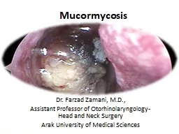
Zamani MD Assistant Professor of OtorhinolaryngologyHead and Neck Surgery Arak University of Medical Sciences Acute invasive fungal rhinosinusitis AIFR is an angioinvasive fungal infection of the nasal cavity and paranasal sinuses that typically develops in ID: 920215

patients aifr commonly fungal aifr patients fungal commonly nasal survival orbital species endoscopic exenteration disease extent surgical surgery role

Share:

Link:

Embed:

Download Presentation from below link

Download Presentation The PPT/PDF document "Mucormycosis Dr. Farzad" is the property of its rightful owner. Permission is granted to download and print the materials on this web site for personal, non-commercial use only, and to display it on your personal computer provided you do not modify the materials and that you retain all copyright notices contained in the materials. By downloading content from our website, you accept the terms of this agreement.


Presentation Transcript

Slide1

Mucormycosis

Dr. Farzad Zamani, M.D.,Assistant Professor of Otorhinolaryngology-Head and Neck SurgeryArak University of Medical Sciences

Slide2

Acute invasive fungal rhinosinusitis

(AIFR) is an angioinvasive fungal infection of the nasal cavity and paranasal sinuses that typically develops in immunocompromised patients, such as those with hematologic malignancies, acquired immunodeficiency syndrome, neutropenia, and diabetes.

Slide3

M

ortality rates of 40% to 80% in the literature.Patients with AIFR present with fevers and localizing symptoms, most commonly facial swelling, nasal congestion, ophthalmoplegia

,

proptosis

, and

vision loss.

Slide4

The most common

organisms: 1. Aspergillus species (most commonly A. fumigatus or A. flavus ) 2. Fungi from the Zygomycetes class, which cause mucormycosis(Rhizopus oryzae)

Slide5

All patients with suspected AIFR should undergo prompt nasal endoscopic evaluation with

particular attention to nasal sensation!

Slide6

Areas of

mucosal pallor, crusting, or necrosis, which are most commonly seen on the middle turbinate should be biopsied and sent for frozen section!

Slide7

The diagnosis can be made with visualization of

necrosis, fungal forms, and angioinvasion on histopathologic analysis.Aspergillus species demonstrate septate hyphae that branch at acute angles (45 degrees) and Zygomycetes display ribbon-like hyphae that are nonseptate and branch irregularly

.

Fungal

cultures

such as

Calcofluor-white and

Grocott

methenamine

silver (GMS)

, as well as

permanent

pathology.

Slide8

Cultures may also reveal less common causative organisms such as Fusarium and

Alternaria species.

Slide9

Imaging studies should be considered as an

adjunct to assess for extent of disease, but should not substitute for a thorough endoscopic examination!CT scans may reveal nonspecific opacification, with more worrisome findings such as bony erosion typically occurring late in the disease process. The role of MRI has been evaluated more recently; it may be a more accurate test for assessment of disease extent due to the

loss of contrast enhancement

seen

in devitalized

mucosa involved by angioinvasion.

Slide10

Management of AIFR

Includes antifungal therapy, surgical resection, and reversal of immunocompromise. Extent of surgery is controversial, particularly with regard to the need for orbital exenteration!Current evidence:There may not be an additional survival benefit provided by orbital exenteration!

Slide11

In patients with

neutropenia, some have suggested a role for granulocyte transfusions, although evidence is limited to small case series.A large systematic review revealed that surgical resection and the use of liposomal amphotericin B were associated with improved survival in patients with AIFR.

Better overall survival

was also associated with

diabetes

.Poorer

overall survival was associated with intracranial involvement and advanced age.

Slide12

Surgical approach:

Endoscopic sinonasal surgery is the mainstay of treatment.Initial biopsy is essentialSurgical debridement is commonly included of turbinectomy (middle and inferior), ethmoidectomy, medial maxillectomy, antrostomy, draf

I-III, and removal of all

devitilized

tissue to find bleeding points in the normal vital tissue!

Slide13

Open approach

Maxiellectomy (partial, subtotal, total, and extended i.e., orbital exenteration) and removal of the palate

Slide14