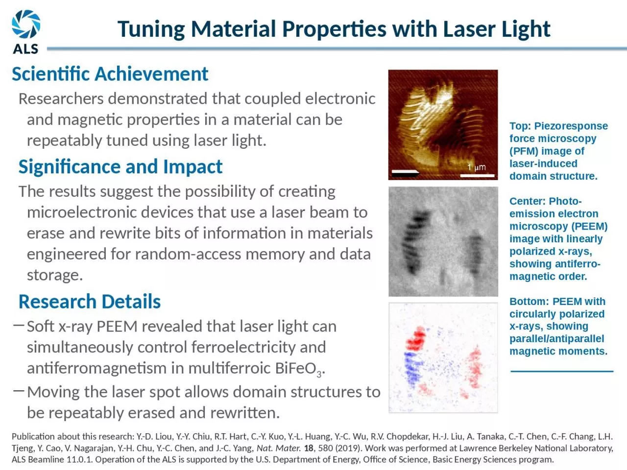
PPT-Top: Piezoresponse force microscopy (PFM) image of laser-induced domain structure.
Author : Outlawking | Published Date : 2022-08-04
Center Photoemission electron microscopy PEEM image with linearly polarized xrays showing antiferromagnetic order Bottom PEEM with circularly polarized xrays showing
Presentation Embed Code
Download Presentation
Download Presentation The PPT/PDF document "Top: Piezoresponse force microscopy (PFM..." is the property of its rightful owner. Permission is granted to download and print the materials on this website for personal, non-commercial use only, and to display it on your personal computer provided you do not modify the materials and that you retain all copyright notices contained in the materials. By downloading content from our website, you accept the terms of this agreement.
Top: Piezoresponse force microscopy (PFM) image of laser-induced domain structure.: Transcript
Center Photoemission electron microscopy PEEM image with linearly polarized xrays showing antiferromagnetic order Bottom PEEM with circularly polarized xrays showing parallelantiparallel magnetic moments. Lecture . 07: . Confocal Microscopy. Adding the Third Dimension. Lecture . 7: Confocal Microscopy. Optical Sectioning: adding the third dimension. Wide-field . Imaging. Point . Spread Function. Deconvolution. Chelsea Aitken. Peter Aspinall. http://www.zyvexlabs.com/EIPBNuG/EIPBN%20images/05Ion.jpg. Intro to Electron Microscopy. Similar to optical microscopy except with electrons rather than photons. Used to image samples with a resolution of 10 Å. Laboratory of Advanced Biological Spectroscopy (L.A.B.S.). University of Milan - Bicocca. Paolo Pozzi. Neuroscience Department. University of Pavia. Flow Cross Correlation in Zebrafish Vascular System. Lecture 18:. High speed microscopy, Part 2. High speed microscopy, Part . 2: Spatial . light modulator microscope and other 3D sensors. Making laser scanning confocal microscopes faster. Resonant scanner confocal. Fire Protection Laboratory Methods Day. June 25, 2014. Paul M. Anderson. Graduate Research Assistant. University of Maryland. Department of Fire Protection Engineering. Transmission Electron Microscopy. Fire Protection Laboratory Methods Day. June 25, 2014. Paul M. Anderson. Graduate Research Assistant. University of Maryland. Department of Fire Protection Engineering. Transmission Electron Microscopy. Jenny . Malmstrom. AFM invented by Binning and co-workers in 1986. . Belongs to the Scanning Probe Microscopy family. Binning et al., Physics Review Letters 1986. AFM invented by Binning and co-workers in 1986. . Lecture . 07: . Confocal Microscopy. Adding the Third Dimension. Andres Collazo, Director Biological Imaging Facility. Wan-. Rong. (Sandy) Wong, Graduate Student, TA. Lecture . 7: Confocal Microscopy. phosphoglucosamine. mutase. Andrew Muenks. 1. , . Tommi. A. White. 1&2. , and Lesa J. Beamer. 1. Department of Biochemistry. 1. and Electron Microscopy Core. 2. , University of Missouri, Columbia, MO. Microscopy is the technical field using microscopes to view samples and objects that can not be seen without unaided eye (objects that are not within the resolution range of the normal eye). There are three well-known branches of microscopy: optical, electron and scanning probe microscopy.. Cantilever-based microscopy in biophysics Clementine Fournier November 15, 2016 Image Credit: Muller et al. (2008) 2 Outline Cantilever-based microscopy in biophysics I. Operating principles of an Atomic Force Microscope (AFM) and Kavantzas N., "Computer vision algorithms in DNA ploidy image analysis", Imaging, Manipulation and Analysis of Biomolecules, Cells and Tissues IV, Proc. SPIE, 6088:60880O (2006) and Loukas S., "A Replica of van 1670 Moody Use the information in this tutorial to supplement the visuals in lab and the Chapters 1 8 and 9 in your lab manual Replica of Culpepper tripod microscope built c 1725 by Co Outline. Introduction. Optical Microscopy. Types of Optical Microscopes. Confocal Microscopy. Laser Scanning Confocal Microscopy. Examples. Optical Microscopy. Optical microscopy is the oldest type of microscope and uses visible light and a system of lenses to create an image of the sample.
Download Document
Here is the link to download the presentation.
"Top: Piezoresponse force microscopy (PFM) image of laser-induced domain structure."The content belongs to its owner. You may download and print it for personal use, without modification, and keep all copyright notices. By downloading, you agree to these terms.
Related Documents