PPT-FLUORESCENCE MICROSCOPY PRESENTED BY
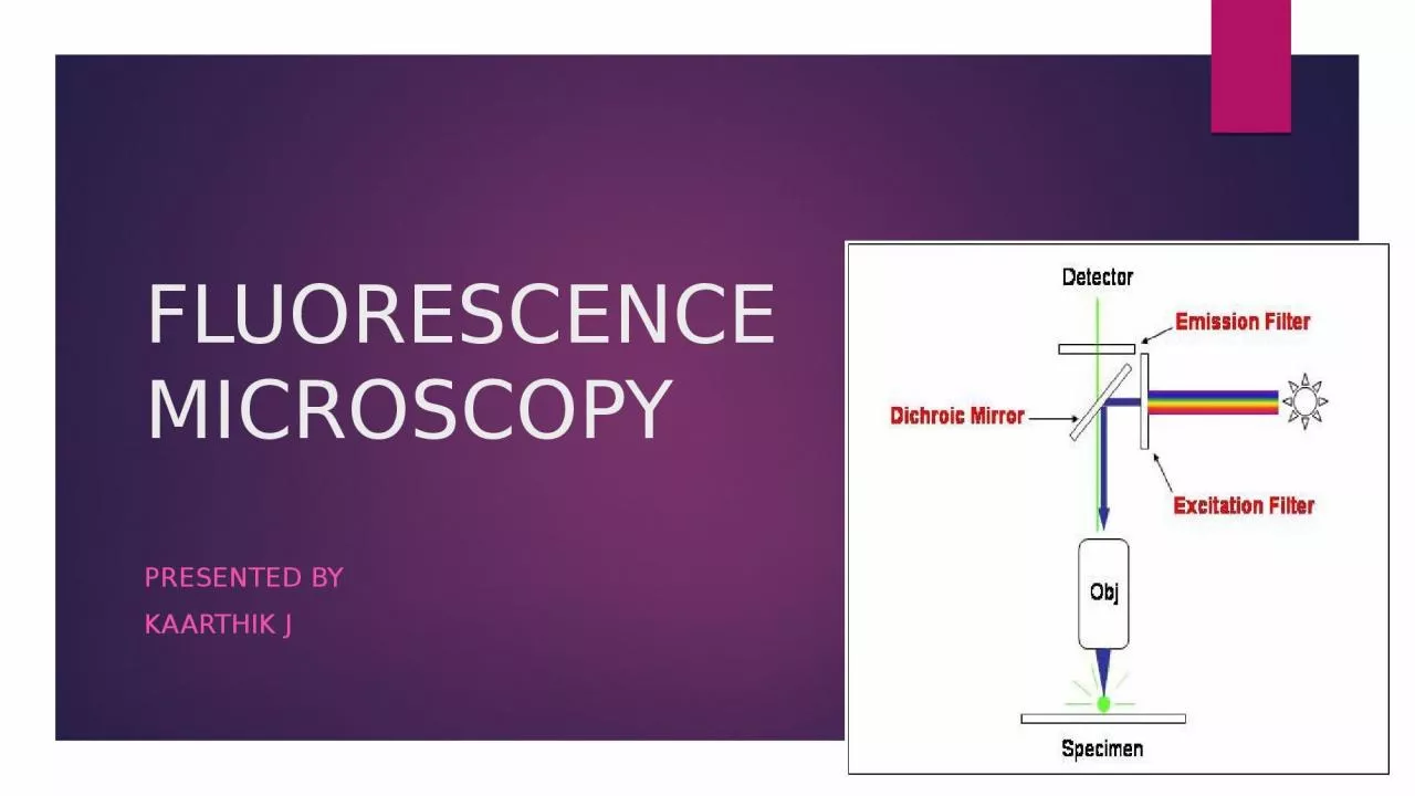
KAARTHIK J INTRODUCTION A fluorescence microscope is a optical microscope that uses fluorescence and phosphorescence instead of or I addition to reflection and absorption
Download Presentation
"FLUORESCENCE MICROSCOPY PRESENTED BY" is the property of its rightful owner. Permission is granted to download and print materials on this website for personal, non-commercial use only, provided you retain all copyright notices. By downloading content from our website, you accept the terms of this agreement.
Presentation Transcript
Transcript not available.