/
complete PLC injury: treatment complete PLC injury: treatment

complete PLC injury: treatment - PowerPoint Presentation

PeacefulPlace
PeacefulPlace . @PeacefulPlace
Follow
342 views
Uploaded On 2022-08-04

complete PLC injury: treatment - PPT Presentation

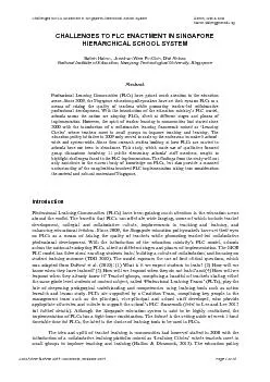
options Trần Đăng Khoa Trương Viết Thông et al BV Chấn thương Chỉnh hình Introduction once referred to as the dark side of the knee occult injuries ID: 935134

plc knee lateral fibular knee plc fibular lateral tibial varus ligament extension larson pcl clinical reconstruction attachment flexion based

Share:

Link:

Embed:

Download Presentation from below link

Download Presentation The PPT/PDF document "complete PLC injury: treatment" is the property of its rightful owner. Permission is granted to download and print the materials on this web site for personal, non-commercial use only, and to display it on your personal computer provided you do not modify the materials and that you retain all copyright notices contained in the materials. By downloading content from our website, you accept the terms of this agreement.


Presentation Transcript

Slide1

complete PLC injury:treatment options

Trần

Đăng

Khoa

Trương

Viết

Thông

et al

BV

Chấn

thương

Chỉnh

hình

Slide2

Introductiononce referred to as the dark side of the knee

occult injuries

associated

with

PCL

or

ACL

tears, with only 28% of

all in

isolation

Slide3

Introductionfail

to address a PLC

injury compromise

concurrent cruciate ligament and lead to early knee degenerative

osteoarthritis

complete PLC lesions treated

surgically

a

cute surgical

intervention results in superior outcomes compared with

chronic reconstruction

Slide4

Anatomy3 major

s

tatic stabilizers:

fibular

c

ollateral

l

igament  popliteus tendonpopliteofibular ligament

Popliteus

COMPLEX

Slide5

Anatomy

Slide6

AnatomyFCL: primary

varus

stabilizer

- femoral attachment:

proximal (1.4 mm) and posterior (3.1 mm) to the lateral

epicondyle

- distal attachment: distal one-third of the lateral aspect of the fibular head

Slide7

AnatomyPLT:

- femoral attachment:

at the point of an average of 18.5 mm anteriorly (17 – 23 mm) from the FCL attachment with the knee at

70

o

-

tibial

attachment: the posteromedial tibia

Slide8

AnatomyPFL

:

- originates

at the

musculotendinous

junction of the

popliteus

- distal attachment: two divisions, the posterior division larger, inserts to fibular head

Slide9

Anatomysecondary stabilizers

: static and dynamic manners

1

/lateral

capsular thickening of the mid-third lateral capsular ligament

2

/coronary

ligament3/lateral gastrocnemius tendon (important landmark)4/fabellofibular ligament

5/long and short head of the bicepsfemoris

6

/

iliotibial

band

Slide10

Anatomy

Slide11

BiomechanicsPLC

:

primary

restraint to

varus

forces as well as

posterolateral

rotationsecondary stabilizer to anterior and posterior tibial

translation

Slide12

BiomechanicsFCL:

primary

restraint to

varus

stress across the

knee

in

regards to tibial external rotation: - FCL and the

popliteus complex are the primary restraints, especially between 30° and 40° of flexion

- PCL is secondary restraint

Slide13

Mechanisms of injurya direct blow to the

anteromedial

knee

hyperextension

non

-contact

varus stress injuriesm

ost often, PLC tears are associated with PCL and/or A

CL

injuries

Slide14

Symptomspain

perceived

side-to-side instability near

extension

difficulty

walking on uneven ground or up and down

stairs

ecchymosisswellingparesthesia

or foot drop; peroneal

nerve

injury occurs

in up to one third of PLC injuries

Slide15

Clinical evaluation

high

index of suspicion

thorough examination

performed bilaterally

Slide16

Clinical evaluationVarus

tress test

: at

0° and 20°

- 30°

knee flexed

20

° - 30: (+)  injury to the FCL and the secondary stabilizers

full

extension:

- (

+)

and restored

injury to the

isolated FCL

- (

+) and

persisted

injury to

the

FCL

,

PLC and cruciate

ligaments

Slide17

Clinical evaluation

Slide18

Clinical evaluationExternal rotational tests

:

d

ial test:

30

0

: (+)

 PLC injury900: (++)

 PLC + PCL injuries

Slide19

Clinical evaluation

Slide20

Clinical evaluationExternal rotational tests

:

Reverse pivot shift test:

(+): the

previously

subluxated

lateral

tibial plateau reduces at approximately 35 ̊ to 40 ̊ of flexion

Slide21

ImagingRadiographs

AP

l

ateral

sunrise view

l

ong standing AP

varus stress (20° of knee flexion) for

the objective diagnosis

Slide22

ImagingMRI

assist

in the diagnosis of acute

lesions

assess

concurrent

injuries

determine the location of the damaged structures

Slide23

Imaging

Slide24

ClassificationGrade I (0-5mm of lateral opening and minimal ligament disruption

)

Grade II

(6-

10mm of lateral opening and moderate ligament disruption

)

Grade III (>10mm of lateral opening and severe ligament disruption and no endpoint)

Slide25

TreatmentPLC physiological

load-sharing

pattern:

LCL represents a larger in situ force near full extension, decreasing with increasing flexion angle of the

knee

popliteus

complex represents a larger in situ force with the knee in flexion than with the knee in extension

Slide26

Treatment

PLC

reconstruction should be performed at same time or prior to ACL or PCL to prevent early cruciate failure

after

cruciate ligament reconstruction, PLC reconstruction is

initiated

Slide27

Treatment2

surgical

techniques: combined

tibial

-fibular-based

techniques and

fibular-based

techniquesLaPrade (tibial-fibular-based):reconstructing

all three major PLC components at each precise insertion siteincreased

technical difficulty and potential

overconstraint

of external and

varus

rotations of the

knee

Slide28

Treatment2

surgical

techniques: combined

tibial

-fibular-based

techniques and

fibular-based techniquesmodified Larson (fibular-based):reproduce

a physiological tension pattern for LCL and PFL using a single ST autograft

advantages

of technical simplicity and reproduction of a more physiological load-sharing

pattern

Slide29

Treatment

Veltri

and

Warren: advocated

reconstruction of PFL and LCL as sufficient to adequately control

posterolateral

instability such as posterior tibial translation and external and varus rotations

Slide30

Treatment“A comparison of modified Larson and 'anatomic'

posterolateral

corner reconstructions in knees with combined PCL and

posterolateral

corner deficiency.”

Apsingi S1,

Nguyen T,

Bull AM, Unwin A, Deehan DJ, Amis AA.Both PLC reconstructions could restore both external rotation and varus laxity to normal

The three-stranded anatomical reconstruction did not perform better than the modified two-strand Larson technique

Slide31

Techniquesmodified

Larson

:

Slide32

Techniquesmodified

Larson

:

Slide33

Techniquesmodified

Larson

:

Less invasive and less technically

demanding

LCL and PFL

secured independently with intended tension at the intended flexion angle of the knee, achieving differential

physiological tension patterns for eachLCL is secured at full extension with 10 N, whereas the PFL is secured at 90° with 10 N

Slide34

Post-op Rehabilitationhinged functional brace

for

3 months

f

irst 2 weeks:

immobilized in extension

ROM exercises using CPM

device from 0° to 90°partial weight-bearing in extension with

gradual progressionfrom

week

4:

ROM exercises >90°

full

weight-

bearing

Slide35

Complications

Arthrofibrosis

Peroneal

nerve injury (15-29%)

Slide36

Thank you!