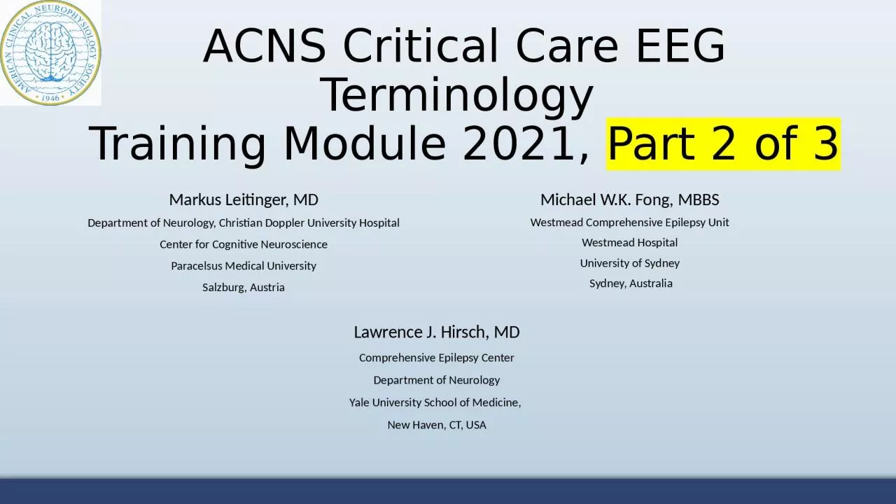
/
ACNS Critical Care EEG Terminology ACNS Critical Care EEG Terminology

ACNS Critical Care EEG Terminology - PowerPoint Presentation

PlayfulPenguin
PlayfulPenguin . @PlayfulPenguin
Follow
381 views
Uploaded On 2022-08-02

ACNS Critical Care EEG Terminology - PPT Presentation

Training Module 2021 Part 2 of 3 Markus Leitinger MD Department of Neurology Christian Doppler University Hospital Center for Cognitive Neuroscience Paracelsus Medical University Salzburg Austria ID: 932337

periodic pattern activity discharges pattern periodic discharges activity wave delta sharp term lateralized pds rhythmic fast rda main spike

Share:

Link:

Embed:

Download Presentation from below link

Download Presentation The PPT/PDF document "ACNS Critical Care EEG Terminology" is the property of its rightful owner. Permission is granted to download and print the materials on this web site for personal, non-commercial use only, and to display it on your personal computer provided you do not modify the materials and that you retain all copyright notices contained in the materials. By downloading content from our website, you accept the terms of this agreement.


Presentation Transcript

Slide1

ACNS Critical Care EEG Terminology

Training Module 2021, Part 2 of 3

Markus Leitinger, MDDepartment of Neurology, Christian Doppler University HospitalCenter for Cognitive NeuroscienceParacelsus Medical UniversitySalzburg, AustriaMichael W.K. Fong, MBBSWestmead Comprehensive Epilepsy UnitWestmead HospitalUniversity of Sydney Sydney, Australia

Lawrence J. Hirsch, MD

Comprehensive Epilepsy Center

Department of Neurology

Yale University School of Medicine,

New Haven, CT, USA

Slide2

Mode of presentation

General remarks:This module has been designed to provide a self guided introduction to the important components of the ACNS standardized critical care EEG terminology, 2021 version. It focuses on discussing terms that are commonly used or with a high clinical importance. It does not describe all of the major and minor modifiers (although it includes many). For comprehensive details on all feaures of the terminology please refer to the ACNS guideline that can be found at

https://www.acns.org/practice/guidelines, under “Continuous EEG Monitoring in Critical Care“, or via the Journal of Clinical Neurophysiology https://journals.lww.com/clinicalneurophys/Fulltext/2021/01000/American_Clinical_Neurophysiology_Society_s.1.aspx

Slide3

Contents, Part 2 of 3

Sequence of topics (follows the full guideline):A. EEG BACKGROUND

B. SPORADIC EPILEPTIFORM DISCHARGESC. RHYTHMIC AND PERIODIC PATTERNS (RPPs)D. ELECTROGRAPHIC AND ELECTROCLINICAL SEIZURES [NEW, 2021]E. BRIEF POTENTIALLY ICTAL RHYTHMIC DISCHARGES (BIRDs) [NEW, 2021]F. ICTAL-INTERICTAL CONTINUUM (IIC) [NEW, 2021]Sequence of slides for each topic:1. Introductory text plus diagram2. EEG example alone with question at the bottom3. EEG with explanatory annotations

4. Summary slide

Slide4

C. Rhythmic and

Periodic Patterns (RPPs)

Slide5

All rhythmic and periodic patterns (RPPs) should be described by main terms 1 and 2, as well as appropriate major and minor modifiers. Not all modifiers will apply to every pattern.

Main term #1 (localization)

(

G

) Generalized

(

L

) Lateralized

(

BI

) Bilateral Independent

(

Mf

) Multifocal

Main term #2 (pattern)

(

PDs

) Periodic Discharges

(RDA) Rhythmic Delta Activity(SW) (Poly)Spike-&-Wave/Sharp- and-Wave

NEW 2021:(UI) Unilateral Independent

RHYTHMIC AND PERIODIC PATTERNS (RPPs): MAIN TERMS

Slide6

RHYTHMIC AND PERIODIC PATTERNS (RPPs): MAIN TERM 2

Main term 2 (pattern description) includes:

PD

: Periodic Discharges

RDA

: Rhythmic Delta Activity

SW

: Spike-and-Wave, Sharp-and-Wave or

Polyspike-and-Wave

Slide7

MAIN TERM 2: PDs

NOTE:

PDs can be blunt

as shown in first example. (

Blunt: having a smooth or sinusoidal morphology.)

Slide8

MAIN TERM 2: RDA

polymorphic, also known as

irregular or arrhythmic

NOT RDA

Slide9

MAIN TERM 2: SW

Spike-and-wave

or Sharp-and-wave (SW): Polyspike, spike or sharp wave consistently followed by a slow wave in a regularly repeating and alternating pattern (spike-wave-spike-wave-spike-wave), with a consistent relationship between the spike (or polyspike or sharp wave) component and the slow wave for at least 6 cycles; and with no interval between one spike-wave complex and the next (if there is an interval, this would qualify as PDs, where each discharge is a spike-and-wave). Longitudinal bipolar

1

2

3

4

5

6

20 to <70 ms

Spike-and-Wave Sharp-and-Wave

1

2

3

4

5

70 to 200 ms

6

duration

measured

at

baseline

duration

measured

at

baseline

Slide10

MAIN TERM 1: GENERALIZED

LEFT

RIGHT

Longitudinal bipolar montage

Generalized: Bilateral synchronous and symmetric periodic discharges. In this case the pattern is “frontally predominant“

Main term 1 (localization) terms have been demonstrated with periodic discharges (PDs).

In practice, any main term 1 can be combined with any main term 2

Slide11

MAIN TERM 1: LATERALIZED

Slide12

MAIN TERM 1: BILATERAL INDEPENDANT

LEFT

RIGHT

Longitudinal bipolar montage

Note: In BIPDs,

lateralized

patterns

occur on each

hemisphere

asynchronously

and at different frequencies

.

Periodic discharges over the left

Independent periodic discharges over the right

Slide13

13

MAIN TERM 1: UNILATERAL INDEPENDANT

Note: In UIPDs, 2 or more lateralized patterns occur within the same hemisphere asynchronously

and at different frequencies

.

Slide14

MAIN TERM 1: MULTIFOCAL

Slide15

In addition to main terms 1 and 2, the following major modifiers can be applied to refine the description of the pattern:

PrevalenceDuration Frequency

PhasesSharpnessVoltageStimulus-Induced (SI-) or Stimulus-Terminated (ST-) (new in 2021)EvolutionPlus (+)

MAJOR MODIFIERS

Slide16

Note that there are 4 distinct time-related scales:

1.

Frequencyrefers to the rate per second or the number of cycles of a pattern within 1 second. Maximum, minimum, and typical frequencies can be recorded. Frequencies are expressed in hertz (Hz) that equates with cycles per second.<0.5, 0.5, 1, 1.5, 2, 2.5, 3, 3.5, and 4 Hz 2. Duration

refers to the typical duration of a single occurrence of the pattern, regardless of whether the pattern occurs rarely or frequently, categorized as: very long, long, intermediate duration, brief, very brief

3.

Prevalence

refers to percent of the entire record occupied by a pattern, categorized as:

continuous, abundant, frequent, occasional, rare

The best estimate of prevalence, when given as a single number, is also known as the

“burden”

of that pattern. E.g., seizure burden of 40%, or IIC burden of 40%, or PD burden of 40%.

4.

Quantification of sporadic

epileptiform discharges

refers to how often sporadic (non-periodic) epileptiform discharges are present in the record, categorized as:

abundant, frequent, occasional, rare16FREQUENCY/DURATION/PREVALENCE

Slide17

What pattern is this?

Slide18

Frontal predominance

110 ms

Sharp morphology

440 ms

DISCHARGE:

<

0.5s,

regardless of phases

10 discharges in

10 seconds

 1 Hz

Generalized Periodic Discharges, GPDs

(frontally predominant, sharp, 1 Hz)

Slide19

Frontal predominance

10 discharges in

10 seconds

 1 Hz

Generalized Periodic Discharges, GPDs

(frontally predominant, sharp, 1 Hz, 3 phases)

110 ms

Sharp morphology

DISCHARGE:

0.5s

BUT

≤ 3 phases

510 ms

3 phases (blue green, orange)

It is still a “discharge“ despite exceeding 0.5s as it has only 3 phases (colored).

Slide20

PERIODIC DISCHARGES: SHARPNESS

Applies to PDs and the S phase of SW

~60 ms

spiky

sharp

~170 ms

1 second

Sharpest phase

:

spiky

(<70 ms

)

Predominant phase

(greatest amplitude):

sharp

(70-200 ms

)

Slide21

Please characterize the pattern

Slide22

LATERALIZED PERIODIC DISCHARGES

Lateralized Periodic Discharges, LPDs

(spiky, with spike-and-wave morphology)

Morphology of one discharge

: spike-and-wave, but

pattern

is LPDs

Despite their spike-and-wave morphology, these are PDs (not SW) as there is a clear interdischarge interval

Slide23

Lateralized Periodic Discharges (LPDs)

Please describe this pattern

Slide24

Lateralized Periodic Discharges (LPDs)

7 EDs/10.5 s= 0.67

0.5-1 Hz

~ 0.37 s

Lateralized Periodic Discharges, LPDs

(right parietal maximum, sharply contoured, positive polarity

,

0.5 - 1 Hz) NOTE: even though the dischages are sharply contoured (i.e., not spiky or sharp), they are still a periodic pattern (LPDs here).

Sharpness: SHARPLY CONTOURED

as duration > 0.2 s

Slide25

Please characterize the pattern

Slide26

Lateralized Periodic Discharges, LPDs

(bilateral asymmetric, 0.5-1 Hz)

LATERALIZED (bilateral asymmetric): bilateral, but clearly and consistently higher amplitude in left hemisphere = LATERALIZED. [Consistently: >80% of the time.]

DISCHARGES:

here: duration ≥500 ms but only one or two phases, therefore still discharges, not bursts.

PERIODIC:

with interdischarge intervals, in green circles

Slide27

Generalized Periodic Discharges (GPDs)

What would you call this pattern?

Slide28

Generalized Periodic Discharges (GPDs)

28

Generalized Periodic Discharges (GPDs),

with shifting asymmetry(bifrontal maximal, sharp, 1.0 Hz)Note that the term “generalized“ allows for higher voltage on one side if there are also discharges with higher voltages on the other side (“shifting predominance“ or more specific “shifting asymmetry“).

If the asymmetry clearly and consistently favors one hemisphere then this would be “lateralized - bilateral asymmetric“; that is not the case here.

higher on LEFT

higher on RIGHT

Slide29

What is the correct term for this pattern?

Reproduced with permission from Hirsch LJ, Fong MWK, Brenner RP. Atlas of EEG in Critical Care, second edition, Wiley: 2022

Slide30

Lateralized Rhythmic Delta Activity (LRDA)

(Left anterior-mid temporal maximum, 1.5 Hz)

Reproduced with permission from Hirsch LJ, Fong MWK, Brenner RP. Atlas of EEG in Critical Care, second edition, Wiley: 2022

Slide31

Reproduced with permission from Hirsch LJ, Fong MWK, Brenner RP. Atlas of EEG in Critical Care, second edition, Wiley: 2022

Please describe the pattern:

Slide32

Reproduced with permission from Hirsch LJ, Fong MWK, Brenner RP. Atlas of EEG in Critical Care, second edition, Wiley: 2022

Polymorphic delta activity (a.k.a. irregular, arrhythmic)

(This is NOT LRDA; RDA has to be rhythmic. This is just focal slowing)

Slide33

Please describe the pattern

Reproduced with permission from Hirsch LJ, Fong MWK, Brenner RP. Atlas of EEG in Critical Care, second edition, Wiley: 2022

Slide34

Lateralized Rhythmic Delta Activity, LRDA

(Right frontocentral maximum, 1.0 Hz)

Reproduced with permission from Hirsch LJ, Fong MWK, Brenner RP. Atlas of EEG in Critical Care, second edition, Wiley: 2022

(Advanced comment:

The fast activity is present even after the RDA ends; thus, this is not +F, just LRDA with no plus. This also means it can not be Extreme Delta Brush pattern.

)

Slide35

What is the correct term for this pattern?

Slide36

Sharp-and-wave:

NO

interdischarge intervals

Generalized sharp-and-wave, GSW

(bifrontal maximum, 1.5 Hz)

17 EDs/10 s

1.5 Hz

90 ms

SHARP

Slide37

What pattern(s) is this?

Slide38

BIPDs

Lateralized Periodic Discharges (LPDs)

38

Bilateral Independent Periodic Discharges BIPDs

Left centroparietal

Right central

Slide39

What is the correct term for this pattern?

Slide40

Unilateral Independant Rhythmic Delta Activity, UIRDA

(Right frontopolar maximum [solid lined box]: 1.5 Hz; Right anterior temporal maximum [dashed box]: 1.0 Hz)

Advanced comment: The fast activity is diffuse and NOT time-locked to the RDA, therefore NO plus and NO delta-brush pattern.

Slide41

What would you call this pattern?

Slide42

12 EDs/10 s

 1.0 Hz5 EDs/10 s

 0.5 Hz

Left frontal

Vertex

Unilateral Independent Periodic Discharges, UIPDs

(orange: maximum left frontal, 1.0 Hz; green: maximum at vertex [Cz], 0.5 Hz)

Focal midline patterns can be deemed in the same

hemisphere (ipsilateral) as an independent pattern in either the left or right hemisphere.

For example, PDs at 1 Hz in the left hemisphere occurring simultaneously with independent focal midline PDs at 0.5 Hz would still qualify as unilateral

independent periodic discharges.

Slide43

Courtesy of

Dr. Luis Octavio Caboclo

This pattern is best classified as:

Slide44

Multifocal Periodic Discharges, MfPDs: 3 independent foci, 1 left, 2 right

Courtesy of

Dr. Luis Octavio CabocloLeft frontal, ~1 Hz

Right frontal, ~0.5 Hz

Right parietal, ~0.3 Hz

Slide45

How often and how long a pattern is present can be described by prevalence and duration:

Prevalence: The percent of the record or epoch that includes the pattern, using the following divisions:

>90% Continuous50-89% Abundant10-49% Frequent1-9% of Occasional

<1% of Rare

45

MAJOR MODIFIERS: PREVALENCE

Slide46

Typical duration of pattern

: The typical duration of a pattern, using the following divisions: (i.e., how long a pattern typically lasts)>

1 hour Very long10-59 min Long [2021 revision, to match ILAE def’n of status epilepticus that is not convulsive (10 min)]1-

9.9 min Intermediate duration

10-59 sec

Brief

<10 sec

Very brief

46

MAJOR MODIFIERS: DURATION

Slide47

The Plus modifier refers to additional features that render the pattern more ictal-appearing than the same pattern without the plus. The Plus modifier can be characterized as:

+F superimposed fast activity

(theta or faster; for PDs or RDA)+R superimposed rhythmic or quasi rhythmic delta activity (for PDs

only)+S superimposed sharp waves/spikes (at least >1/10s but not periodic and not spike-and-wave, SW) or

sharply contoured

(for

RDA

only)

+FR

if both subtypes apply (for

PDs

only)

+FS

if both subtypes apply (for

RDA

only)

“PLUS” MODIFIER

Slide48

Longitudinal bipolar

fast activity associated with each discharge

EXAMPLE A: PDs+F

code as +F if the fast activity is part of the RDA or PD pattern and not simply part of the background activity

Longitudinal bipolar

4

6

5

EXAMPLE B: PDs+F

NOTE:

fast activity cycling with the periodic discharge

PERIODIC DISCHARGES PLUS FAST (

PDs+F

)

OR …

Slide49

code as +F if the fast activity is part of the RDA or PD pattern and not simply part of the background activity

Longitudinal bipolar

EXAMPLE C: PDs+FEXAMPLE D: PDs (NOT +F, as fast activity is part of the background and present even when the pattern is not)

4

6

5

Longitudinal bipolar

1

2

3

6

...

PERIODIC DISCHARGES PLUS FAST (

PDs+F

)

Slide50

PERIODIC DISCHARGES PLUS RDA (

PDs+R)

Slide51

RHYTHMIC DELTA ACTIVITY PLUS SPIKES or SHARP WAVES (RDA+S)

Rhythmic delta activity (in this case over the left hemisphere) with associated spikes.

NOTE the discharges are not periodic (compared to example A, prior slide). If a pattern qualifies as PDs+R and RDA+S it is suggested that it be classified as PDs+R. If the spike/sharp component forms a stereotyped relationship with each wave and there is no inter-discharge interval it would likely qualify as SW (example B, prior slide)LEFT

Slide52

Spiky LPDs+F

52

Please describe the pattern

Slide53

Spiky LPDs+F

53

Lateralized Periodic Discharges with superimposed fast activity, LPD+F

(Lateralized: bilateral asymmetric, right temporal maximum, 1 Hz)

Fast activity:

Artifact

Slide54

What is the best way to describe this pattern?

Slide55

Left LRDA, 1.5-2 Hz

Lateralized Rhythmic Delta Activity, LRDA+S

(it`s +S due to the sharp waves shown by the asterisks)

Slide56

56

What is the correct term of this pattern?

Slide57

57

Lateralized Periodic Discharges with superimposed Rhythmic Delta Activity: LPD+R

(Lateralized: bilateral asymmetric, spiky and sharp, 1.5 Hz)

Sharpness: spiky

Sharpness: sharp

Rhythmic activity

Not LSW since sharp waves are not time locked to delta waves for 6 or more cycles, but close

Slide58

Please describe the pattern

Slide59

Lateralized Periodic Discharges + R (Ictal-Interictal Continuum, IIC)

1.5-2 Hz LPD+R, therefore on the ictal-interictal continuum (discussed later); not >2.5 Hz and not evolving, so does not qualify as a definite electrographic seizure, even though there is a reasonable chance it could cause symptoms or neuronal injury.

24 discharges in

14 seconds

1.5 - 2 Hz

Rhythmic activity

Slide60

EXTREME DELTA BRUSH (EDB)

“Extreme Delta Brush (EDB)”:

A specific subtype of +F:Definite EDB: Consists of either abundant or continuous:A. RDA+F, in which the fast activity has a stereotyped relationship to the delta wave (e.g., always maximal on the upstroke, crest, or downstroke of the wave); ORB. PDs+F, in which each PD consists of a single blunt delta wave with superimposed fast activity, and in which

the fast activity has a stereotyped relationship to the delta wave (i.e., periodic delta brushes)

Possible

EDB

:

Satisfying criterion A) or B) above

EXCEPT

either:

i.

only occasional or frequent (rather than abundant or

continuous

)

OR

ii.

the superimposed fast activity lacks a stereotyped relationship to the delta wave; continuous, invariant fast

activity during RDA would fall into this category

Slide61

Slide62

EXTREME DELTA BRUSH (EDB)

Slide63

EXTREME DELTA BRUSH (EDB)

This is not +F since the fast activity is present even when the RDA is not.

Slide64

What is the correct term for this pattern?

Slide65

Generalized Periodic Discharges with superimposed fast activity (GPD+F)

(Bifrontal maximum, 0.5 Hz): If abundant/ continuous, this is definite EDB; if it‘s frequent/occasional, it is possible EDB.

Periodic blunt delta waves with superimposed beta in a stereotyped relationship to the delta waves.

Slide66

Evolving and fluctuating refer to changes in at least one of the following:

Frequency

Location Morphology(not amplitude alone)If neither term applies, report as static

.

66

MAJOR MODIFIERS: EVOLVING, FLUCTUATING or STATIC

Slide67

At least 2 unequivocal, sequential changes in frequency, morphology or location defined as follows:

Frequency:

≥2 consecutive increases or decreases of ≥0.5 Hz, (e.g., 2  2.5 to 3 Hz, or 3  2 to 1.5 Hz)Morphology:

≥2 consecutive changes to a novel morphologyLocation: sequentially spreading into or out of ≥2 different standard 10-20 electrode locations

NOTE:

if evolving and

>

10s: it’s a seizure.

If evolving, 0.5-10s and reaches >4 Hz, it’s a BIRD (definite due to evolution)

If evolving, <10s and never >4 Hz, just use the modifier “evolving”, e.g., “very brief evolving RDA”

MAJOR MODIFIERS: EVOLVING

Slide68

EVOLUTION OF FREQUENCY

At least 2 unequivocal, sequential changes in frequency defined as follows: Evolution in

frequency is defined as at least 2 consecutive changes in the same direction by at least 0.5 Hz. In order to qualify as present, a single frequency must persist for at least 3 cycles. The criteria for evolution must be reached without the evolving feature (frequency) remaining unchanged for 5 or more continuous minutes.

Slide69

69

At least 2 consecutive changes to a novel morphology.

EVOLUTION OF LOCATION

Evolution in location is defined as sequentially spreading into or sequentially out of at least two different standard 10-20 electrode locations. The two consecutive changes must be in the same category (location) to qualify. In order to qualify as present, a single location must persist for at least 3 cycles.

Slide70

70

At least 2 consecutive changes to a novel morphology.

EVOLUTION OF LOCATION

Slide71

At least 3 changes, <1 min apart, in:

Frequency

(by ≥0.5/s), Morphology, or Location

(by ≥1 standard inter-electrode distance),

BUT

not qualifying as evolving

.

71

MAJOR MODIFIERS: FLUCTUATING

Slide72

FLUCTUATION in FREQUENCY

>

3 changes, not more than one minute apart, in frequency (by at least 0.5 Hz), but not qualifying as evolving. This includes patterns fluctuating from 1 to 1.5 to 1 to 1.5 Hz. In order to qualify as present, a single frequency must persist at least 3 cycles (e.g., 1 Hz for 3 s, or 3 Hz for 1 s).

Slide73

Please describe the pattern

Slide74

74

Lateralized Periodic Discharges, fluctuating.

This EEG is shown on a compressed time scale to demonstrate LPDs slowing from 1 Hz to 0.5 Hz for <1 minute (1st change) and then accelerating back to 1 Hz (2nd change). This patient had fluctuating LPDs but in order to qualify as fluctuation there would have to be at least 3 changes not more than 1 min apart (3rd change not shown). 1

st change (1 Hz to 0.5 Hz)

2

nd

change (0.5 Hz to 1 Hz)

1 Hz

0.5 Hz

1 Hz

<

1

min

<

1

min

Slide75

FLUCTUATION in LOCATION

>

3 changes, not more than one minute apart, in location (by at least 1 standard inter-electrode distance), but not qualifying as evolving. This includes patterns spreading in and out of a single electrode repeatedly. In order to qualify as present, a single location must persist at least 3 cycles.

Slide76

FLUCTUATION in LOCATION

Slide77

Many patterns including seizures can be reproducibly brought about or exacerbated by an alerting stimulus, with or without clinical alerting, when a patient is in their less-stimulated state. The phenomenon was initially described as stimulus-induced rhythmic, periodic, or ictal appearing discharges (SIRPIDs). Since then, it has become evident that the content of the stimulus-induced pattern, rather than the fact that it is stimulus induced or not, carries the majority of significance. It is therefore encouraged to apply the term stimulus induced (SI-) or stimulus terminated (ST-) to the respective RPPs or seizures, as opposed to referring to them as a collective

. Patterns can be classified as:

Stimulus induced (SI-)Stimulus terminated (ST-)SpontaneousUnknown, including unclear and untested

MAJOR MODIFIERS: STIMULUS INDUCED/ TERMINATED

Slide78

Describe the EEG changes following stimulation with sternal rub at the time of the red bar

Slide79

Stimulus Induced Generalized Periodic Discharges, SI-GPD

(0.5 Hz)

Slide80

SIRPIDs: example 2B

Reproduced with permission from

Hirsch LJ, et al. Epilepsia 2008 Please classify the EEG; intranasal tickle was performed in the middle of part A

Slide81

SIRPIDs: example 2B

Stimulus-Induced Seizure (electroclinical sz.), SI-Seizure:

STIM in part A (first arrow) leads to gradual development of a bilateral evolving ictal pattern, bitemporal maximum, over the next 30 secs (B-D), evolution in frequency: 10 hz 7 Hz  4 Hz [end of sz not shown]; clinical correlate: mouth tightened, then finger twitching in left hand only, then eyes opened wide and deviated up and to the right. Recurred whenever stimulated.

Reproduced with permission from Hirsch LJ, et al. Epilepsia. 2008

Slide82

END OF PART 2

CONTINUE

TO PART 3