/
Skin Physiology 101 The Global Beauty Group Skin Physiology 101 The Global Beauty Group

Skin Physiology 101 The Global Beauty Group - PowerPoint Presentation

Powerpixel
Powerpixel . @Powerpixel
Follow
344 views
Uploaded On 2022-08-01

Skin Physiology 101 The Global Beauty Group - PPT Presentation

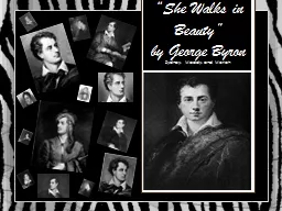
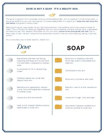
Knowledge of the structure functions and conditions of the skin are essentials for understanding how to optimally serve our clients at every level Did you know The skin is the bodys largest organ ID: 931766

layer skin dermis cells skin layer cells dermis body collagen epidermis responsible stratum layers blood free vessels elastin fibers

Share:

Link:

Embed:

Download Presentation from below link

Download Presentation The PPT/PDF document "Skin Physiology 101 The Global Beauty Gr..." is the property of its rightful owner. Permission is granted to download and print the materials on this web site for personal, non-commercial use only, and to display it on your personal computer provided you do not modify the materials and that you retain all copyright notices contained in the materials. By downloading content from our website, you accept the terms of this agreement.


Presentation Transcript

Slide1

Skin Physiology 101

The Global Beauty Group

Knowledge of the structure, functions and conditions of the skin, are

essentials for understanding how to optimally serve our clients at every level

Slide2

Did you know…

The skin is the body’s largest organ

It has 3 layers: the

epidermis, dermis and the subcutaneous layerIt accounts for approximately 15% of our body weight and the average person has roughly 300 million skin cells.Flattened, the average persons skin covers 2 square metresIt is the most sensitive organ of the human body and has at least 5 different types of receptors that respond to pain & touch.The conventional body system term for the skin is the Integumentary System, which includes the hair and nails as well, however the skin alone is referred to as the cutaneous membraneThe skin is the thickest on the palms of the hands and the soles of the feet; and the thinnest on the eyelids.1 square cm of skin contains: 15 sebum glands (sebaceous)700 sweat glands (sudoriferous) Almost 1 metre of blood vesselsapproximately 3.5 m of nerves200 nerve endings10 hairs and 3 million cells of which, 3000 are sensory cells (Body structures & Functions 13th Edition, Ann Scott & Elizabeth Fong, 2016)

Slide3

Skin Functions

P

rotection,

Sensation, Heat Regulation, Excretion, Secretion, Absorption, Vitamin D Synthesis

Slide4

Skin Functions

Protection

The skin acts as a protective barrier to outside elements and microorganisms. It protects underlying tissues from microbes, abrasion, heat & chemicals. It prevents dehydration and shields against the effects of UV rays

Blood vessels protect the body from invasion of toxins and microbes by sending out “combat-unit” cells, known as Leukocytes.SensationSensory nerve endings in the dermis sense touch, pain, cold, heat and pressure and serve as body’s communication in perception and interaction. Hairs also act as touch receptors due to the way that nerve endings wrap around the base of the follicle.

Slide5

Skin Functions

Heat regulation (aka-Thermoregulation)

The body’s average temperature sits at 37°C. In order to maintain homeostasis, the skin will adjust itself to warm or cool the body.

Hair follicles and sweat glands disperse heat to keep us from over-heating as the evaporation of sweat from the skins surface helps to lower body temperature. The body can also cool itself down via vasodilation. When the vessels in the dermis dilate, more blood flows through the dermis which increases the amount of heat loss from the body. On the other hand, our cutaneous blood vessels constrict when we are cold, decreasing the blood flow to the skin to allow for warm blood to circulate our vital organs. ExcretionSweat glands (sudoriferous glands) excrete perspiration and detoxify the body of salts, carbon dioxide, ammonia & urea.

Slide6

Skin Functions

Secretion

Oil (

sebum) protects the surface of the skin, and lubricates skin and hair. Sebum is created to soften skin, slowing down H2O evaporation (also known as trans epidermal water loss (TEWL)).Sebum secretion is controlled by the hormonal system, meaning there is increased production during puberty and pregnancy and decreased production with old age. It also contains antibacterial and anti fungal properties.AbsorptionVitamins, Carbon dioxide (CO2), Oxygen (O2), chemicals and topical steroids and creams can all penetrate the skin, for better or worse.Vitamin D synthesis Vitamin D synthesis aids in the absorption of calcium and increases the production of antimicrobial substances that regulate immune function and help to reduce inflammationOnly small amounts of UV exposure are required for the synthesis of Vitamin D (10-15 minutes, twice weekly)

Slide7

Layers Of The Skin

Slide8

Layers Of The Skin

Epidermis

The epidermis is the most superficial layer of the skin. It predominantly consists of

keratinocytes and is made up of 5 separate layers of it’s own (Stratum Germinativum or Stratum Basale, Stratum Spinosum, Stratum Granulosum, Stratum Lucidum and Stratum Corneum).It is responsible for protection, cellular regeneration and melanin (pigment) production.The epidermis has regular cellular turnover due to the constant ‘shedding’ and regeneration of skin cells. The average epidermis is approximately 0.1mm thick . This means that the average epithelial cell (in young, healthy skin) will take roughly 28 days to move from the basal layer of the epidermis to the most superficial layer. As we age, the rate of cellular turnover slows, and this life cycle can take as long as 42+ days.As epidermis does not contain any blood vessels, it relies on the structures of the dermis for nutrient delivery and waste disposal.Pigment producing cells, better known as melanocytes, are also located in the basal layer of our epidermis

Slide9

Layers Of The Skin

Dermis

The dermis is the middle layer of the skin and houses blood vessels, lymphatic vessels, hair follicles, connective tissue, sebaceous & suderiforous glands, nerves and additional skin appendages

It is the ‘live-layer’ often referred to as the ‘true skin’ and is approximately 5-7 times thicker than the epidermis It consists of 2 layers of it’s own, commonly known as the papillary and reticular dermis. The papillary dermis is thinner and sits superficially in comparison to the reticular dermis. It is responsible for providing the epidermis with essential nutrients and also assists in temperature regulationThe reticular dermis is thicker and contains dense connective tissue responsible for supporting the skinSubcutaneous Also known as the Hypodermis, this layer of the skin is the deepest layer. It sits between the dermis and underlying tissues and organs and is predominantly made up of fatty tissue (adipose). This is where most body fat is stored. It is highly elastic and fat cells act as a ‘shock absorber’ to support the delicate structures of the skin

Slide10

The Epidermis

Stratum

Corneum

Stratum LucidumStratum GranulosumStratum SpinosumStratum Germinativum

Slide11

5 Layers Of The Epidermis

Stratum

Corneum

(25-30 layers)Also referred to as the “horny layer”It is the outer most layer of the skin and is therefore the only layer that can be seen with the human eye. The Stratum Corneum is made up of dead skin cells containing keratin and have no nucelus.The cells in this layer serve as a protective barrier against environmental trauma including heat, light, physical abrasion, chemicals and microorganismsIt also acts as our waterproof layerStratum Lucidum (4-6 layers)This is the transparent layer, found only in the ‘thick skin’ on the soles of the feet and palms of the hands,Also consists of dead skin cells due to their distal location from the blood supply under the basal layer

Slide12

5 Layers Of The Epidermis

Stratum Granulosum (3-5 layers)

A layer of granular-looking cells which are ‘dying’ and on their way up to replace the existing Stratum

Corneum. The nuclei of the cells in this layer are in various stages of degeneration. As they break down, these cells can no longer carry on vital metabolic reactions and as a result, they die.Large quantities of keratin/protein are created in this layer to act as protection for the lower layers of the skin.Organelles within these keratinocytes are responsible for the release of the lipid-rich secretion that is deposited between the cells of the granulosum, lucidum and corneum that acts as a water repellant sealant. Stratum Spinosum (8-10 Layers)Also referred to as the prickle cell layer or the spiny layer due to the intercellular connections (also known as desmosomes) resembling little ‘spines’ which anchor cells together and provide supportThe Langerhans cells are located in this layer. These are responsible for assisting the lymphocytes in protecting the body from infection. They act as the immune system’s radar to alert immune cells to an invasion or attack

Slide13

5 Layers Of The Epidermis

Stratum Germinativum (1 layer)

Is also referred to as the basal layer and is made up of a single layer of

cuboidal cells. This is the only layer of the epidermis that can regenerate through a process called mitosis. It is responsible for the growth of new skin cells via a process of cell division. This is also where our melanocytes are located. Melanocytes are responsible for creating our skins pigment, also known as melanin. The role of melanin is to protect the skin from the harmful effects of ultra violet radiation. Melanin is transported via dendritic arms from the Stratum Germinativum to the stratum spinosum where it forms a ‘cap’ over the cells. This is our body’s natural defense mechanism and helps to protect our skin from UV induced cell destruction. Approximately 7% of skin cells are melanocytes. Everyone has the same number of melanocytes, meaning that skin colour is not determined by melanocyte quantity, but instead by melanocyte activity. The more melanin your melanocytes produce, the darker your skin colour (measured on the Fitzpatrick scale)

Slide14

Fitzpatrick Skin Types

Skin Type

Skin Color

CharacteristicsIWhite; very fair; red or blond hair; blue eyes; freckles

Always burns, never tans

II

White; fair; red or blond hair; blue, hazel, or green eyes

Usually burns, tans with difficulty

III

Cream white; fair with any eye or hair color; very common

Sometimes mild burn, gradually tans

IV

Brown; typical Mediterranean caucasian skin

Rarely burns, tans with ease

V

Dark Brown; mid-eastern skin types

V

ery rarely burns, tans very easily

VI

Black

Never burns, tans very easily

Slide15

The Dermis

Papillary Dermis

Reticular Dermis

Slide16

2 Layers Of The Dermis

Papillary Dermis (Papillary Layer)

The papillary layer makes up approximately 1/5

th of the total thickness of the dermisIt sits directly underneath the Stratum Germinativum and houses the capillaries that nourish the epidermisThis portion does contain collagen and elastin fibers however the collagen fibers are thin and the elastin fibers are fineThe papillary dermis also houses nerve endings that sense touch, as well as free nerve endings that help sense warmth, cold, pain, tickling and itching

Slide17

2 Layers Of The Dermis

Reticular Dermis (Reticular Layer)

The reticular layer makes up the remaining 4/5

th of the total thickness of the dermisIt sits in between the papillary layer of the dermis and the subcutaneous layer of the skinAids in the removal of carbon dioxide and waste products.Blood vessels within this layer assist in the transportation of O2 from the lungs to all of the body’s cells for cell respiration and metabolism which also nourishes the skin.Collagen and elastin are created by specialized cells called fibroblasts. The collagen fibers in this layer are thicker than the collagen fibers found in the papillary layer and their arrangement is more regular. Elastin fibers in this layer are also coarse. The improved arrangement of collagen and elastin fibers provide better structure. This is what gives the skin its strength, extensibility and elasticity. It gives it the ability to stretch and return to its original shape The space between the fibers of the dermis contain hair follicles, nerves, sebaceous glands, sudoriferous glands and small amounts of adipose cells in the deeper layerThe reticular dermis also contains scattered fibroblasts and macrophagesNote: the dermal blood vessels are so extensive, they hold between 5-9% of all of the blood in the body

Slide18

Subcutaneous Layer

Also referred to as the Hypodermis or adipose tissue, this layer is

responsible for connecting the dermis to the muscles and bones, providing insulation and thermoregulation and protecting muscles and bones from impact through shock absorption. It also assists the function of the dermis by providing support to the blood vessels, lymphatic vessels, nerves, and glands that pass through it to reach the dermis.

It constantly accumulates and stores fat cells, commonly known as adipocytes, which can also act an energy reserve. The number of adipocytes in the subcutaneous tissue varies with the area of the body, however the size of the individual cells depends on the individual.

Slide19

Subcutaneous Layer

Other components that can be found in the Subcutaneous layer include:

Fibrous bands: to anchor the skin to the deep fascia

Collagen and elastin fibers: to connect the subcutaneous tissue to the dermisLymphatic vessels: to connect the dermis to the lymphatic systemNerves: to connect the dermis and free nerve endings in the subcutaneous tissue to the nervous systemBlood vessels: to connect the dermis to the circulation of bloodBursae: to facilitate the smooth movement of skin over joints in the bodyPanniculus carnosus: to allow skin to move independently of the deeper muscle massMast cells

Slide20

The extracellular matrix (ECM) is an active component of all living tissues. It provides structural support and facilitates cell division, growth, and development. It largely determines how a tissue looks and functions

The extracellular matrix is comprised of specialised protein molecules, water, minerals, and fibrous proteins.

The components of the extracellular matrix are made and organised by the cells that live within it. In most tissues, 

fibroblasts, are largely responsible for this.Fibroblasts are one of the most prevalent cells in the dermis. They are located in both the dermal papillae and the reticular dermis. Their main function is to maintain skin integrity through synthesis of ECM proteins such as collagen, elastin, glycosaminoglycans, and glycoproteins. The structural composition varies depending on their location in the body however fibroblast proliferation is stimulated as a result of injury, which in turn accelerates the wound healing response through the production of proteins responsible for healing and regeneration. The Extracellular MatrixFibroblast

Slide21

Whilst fibroblasts are the most prevalent

cell

in the dermis, the most predominant

molecules in the extracellular matrix are proteoglycans and fibrous proteinsProteoglycans Proteoglycans form when glycosaminoglycan (GAG) chains bind to core proteins. There are several classes of GAGs and their chemical properties are determined by the types of sugars found in their polysaccharide chains. GAGs are negatively charged hydrophilic molecules. They have the ability to absorb and store large amounts of water due to the strong attraction. One example is Hyaluronic AcidFibrous proteins (collagen, elastin, fibronectin, and laminin)These are found in varying amounts within the extracellular matrix of different tissues. These proteins are produced by fibroblasts.Collagen: strong, stretch-resistant fibre that provides tensile strength to your tissues. It's the most abundant protein in the human body. Elastin: stretchy and resilient protein. Much like a rubber band, elastin permits tissues to return to their original shape after they've been stretched.Fibronectin: helps position cells within the extracellular matrix.Laminin: forms sheet-like networks that serve as the 'glue' between dissimilar tissues.The Extracellular Matrix

Slide22

(A)

Collagen is a triple helix formed by three extended protein chains that wrap around one another. Many rod like collagen molecules are cross-linked together in the extracellular space to form collagen fibrils (top) that have the tensile strength of steel. The striping on the collagen fibril is caused by the regular repeating arrangement

of the collagen molecules within the fibril.

(B) Elastin polypeptide chains are cross-linked together to form rubberlike, elastic fibers. Each elastin molecule uncoils into a more extended conformation when the fiber is stretched and will recoil spontaneously as soon as the stretching force is relaxed. Structure of Collagen & Elastin

Slide23

Structure of Collagen & Elastin

Slide24

Types of Ageing

There are two distinct types of ageing:

Intrinsic aging is

genetically determined and inalterable. It is not constant across different populations or even different anatomical sites on the same individual.Extrinsic aging is determined by external and environmental factors such as sun exposure and lifestyle (smoking, drinking etc.). Scientist have determined that 85% of all aging is extrinsic.

Slide25

Factors affecting intrinsic skin ageing:

Ethnicity

Anatomical variations

Hormonal changes Illness or other bio chemical changesGravity and time on earth!Factors affecting extrinsic ageing:Lifestyle Influences (temperature and humidity)MedicationSmokingExposure to UVBad health habits (i.e. poor nutrition, smoking, drinking).Poor NutritionLack of proper skin care -impaired barrier function due to lack of hydration, proper protection antioxidants (aka cell protectants) sunscreens.Types of Ageing

Slide26

Free Radicals

Humans depend on oxygen to produce the energy required for cells to carry out their normal functions.

A cell’s mitochondria (also referred to as the engine of the cell) is responsible for the conversion of oxygen into energy. But this process also leaves a kind of exhaust product known as free radicals.

As the body ages, it produces more and more free radicals. This results in the generation of highly reactive oxygen molecules that inflict cellular damage by reacting with biomolecules including DNA, proteins, and lipids. When free radicals are not destroyed by antioxidants, they create cellular degeneration (or oxidative stress) by means of a process known as oxidation. Just like rust or a sliced apple turning brown, we are subject to that same process and are more vulnerable to it as we age. A lifetime of oxidative stress leads to general cellular deterioration associated with aging and degenerative diseases.

Slide27

Free Radicals

Free radicals are also produced by sunlight, toxins such as pesticides, cigarette smoke and air pollution. They are without question the central players in the ageing process.

Because electrons normally come in pairs, the free radicals collide with other molecules in an attempt to steal an electron, and may start a chain reaction, damaging your DNA and cells.

Free radicals also cause harm to lipids and proteins. Their damaging potential may be counteracted by antioxidants

Slide28

Antioxidants

Antioxidants can be vitamins, amino acids (proteins), and other natural substances that help the cells to cope with the effects of the environment.

Nature knows…and has provided us with an abundance of protective fruits, vegetables, grains, and cereals which are rich in the antioxidants.

Antioxidants neutralize free radicals, which if left to their own devices, will destroy cells.“Antioxidants scoop up free radicals , preventing the cellular degeneration and production of chemicals within the body that cause further damage.” (Nikolas Perricone, M.D.)Visualize antioxidants as nature's demolition teams that neutralize the free radicals before they can do any damage. They are any substances that prevent or slow the oxidation process and work by donating an electron to a free radical so it becomes a stable oxygen molecule. Free radicals cause oxidation - and antioxidants prevent oxidation.

Slide29

Glossary

Epidermis

-

the thin, outer layer of the skin that is visible to the eye. Doesn’t contain any blood vessels and is therefore dependent on the dermis for nutrient delivery and waste disposal.Dermis - the layer of skin that lies beneath the epidermis and above the subcutaneous layer. Consisting of the papillary and reticular layers, It is the thickest layer of the skin, and is made up of fibrous and elastic tissue. It provides the skin with strength and flexibilityPapillary Dermis - the superficial layer of the dermis laying just beneath the epidermal junction. It is relatively thin and is made up of loose connective tissue including capillaries, elastic fibers, reticular fibers, collagenReticular Dermis - the deeper and thicker layer of the dermis, which lies above the subcutaneous layer of the skin. It contains dense connective tissue including blood vessels, elastic fibers, collagen fibers, fibroblasts, mast cells, nerve endings and lymphaticsSubcutaneous layer - also known as the hypodermis and is the innermost layer of skin. It's made up of fat and connective tissues that house larger blood vessels and nerves. It acts as an insulator and regulates body temperatureHomeostasis - the ability of the body to maintain a stable internal environment despite changes in external conditionsLeukocytes - part of the body's immune system. They help the body fight infection and other diseases.Sudoriforous glands (Sweat Glands) – found in the dermis and are responsible for the production of sweat in response to certain conditions, such as heat and stress. As the sweat evaporates off the skin, it cools the body down to maintain homeostasisSebaceous glands (Oil Glands) – also found in the dermis and are responsible for the secretion of sebum, an oily substance that helps to keep the skin moist and helps to prevent the entry of foreign substancesSebum - a light yellow, oily substance that helps to keep the skin and hair moisturized. Sebum is made up of triglycerides, free fatty acids, wax esters, squalene, cholesterol esters, and cholesterolKeratinocytes - responsible for the synthesis of the protein keratin. Originate from the deepest layer of the epidermis, and gradually move up to the outside layer of the epidermis. Here they are shed from the skin and replaced by new maturing cellsMelanocytes - responsible for the production of melanin, which contributes to the colour of our skin. It also helps to protect the body from ultraviolet radiation present in sunlight that can damage the DNA of the skin cellsMelanin - responsible for determining skin and hair colour and is present in the skin to varying degrees

Langherhans

cells -

work to detect foreign substances and infections, as a part of the immune system of the skin.

Mitosis -


a process of cell division whereby a cell divides to form two identical cells. The major purpose of mitosis is for growth and to replace worn out cells

Extracellular matrix -

a meshwork of proteins and carbohydrates that provides a structural support scaffold for cells. Also responsible for controlling

communication between cells, segregating tissues and regulating cell processes such as growth, migration and differentiation.

Fibroblasts -

responsible for making the extracellular matrix and collagen. Forms the structural framework of tissues and plays an important role in tissue repair. Fibroblasts are the main connective tissue cells present in the body

Slide30

Thank you!