/
In vitro evaluation and in vivo efficacy of In vitro evaluation and in vivo efficacy of

In vitro evaluation and in vivo efficacy of - PowerPoint Presentation

Ruggedman
Ruggedman . @Ruggedman
Follow
342 views
Uploaded On 2022-08-04

In vitro evaluation and in vivo efficacy of - PPT Presentation

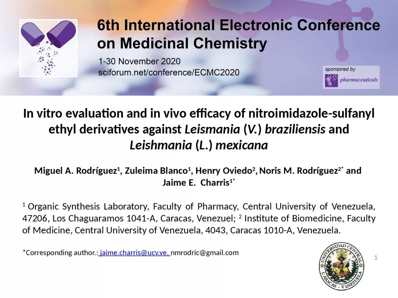
nitroimidazolesulfanyl ethyl derivatives against Leismania V braziliensis and Leishmania L mexicana Miguel A Rodríguez 1 Zuleima Blanco 1 Henry Oviedo 2 ID: 935176

braziliensis compounds results mexicana compounds braziliensis mexicana results leishmaniasis promastigotes venezuela treated discussion infected mice fig 2eq biological activity

Share:

Link:

Embed:

Download Presentation from below link

Download Presentation The PPT/PDF document "In vitro evaluation and in vivo efficacy..." is the property of its rightful owner. Permission is granted to download and print the materials on this web site for personal, non-commercial use only, and to display it on your personal computer provided you do not modify the materials and that you retain all copyright notices contained in the materials. By downloading content from our website, you accept the terms of this agreement.


Presentation Transcript

Slide1

In vitro evaluation and in vivo efficacy of nitroimidazole-sulfanyl ethyl derivatives against Leismania (V.) braziliensis and Leishmania (L.) mexicanaMiguel A. Rodríguez1, Zuleima Blanco1, Henry Oviedo2, Noris M. Rodríguez2* and Jaime E. Charris1*1 Organic Synthesis Laboratory, Faculty of Pharmacy, Central University of Venezuela, 47206, Los Chaguaramos 1041-A, Caracas, Venezuel; 2 Institute of Biomedicine, Faculty of Medicine, Central University of Venezuela, 4043, Caracas 1010-A, Venezuela.*Corresponding author.: jaime.charris@ucv.ve. nmrodric@gmail.com

1

Slide2

Graphical Abstract In vitro evaluation and in vivo efficacy of nitroimidazole-sulfanyl ethyl derivatives against Leismania (V.) braziliensis and Leishmania (L.) mexicana2

Metronidazole

:

Drug

Repositioning

Optimization

against

Leishmania

spp

.

L. (V)

braziliensis

DL50 21

ug

/

mLL. (L) mexicana DL50 18 ug/mL

Mice

infected with Leishmania species:Reduced toxicity Less size of lesionsLonger survival time

Slide3

Abstract:The aim of this study was the synthesis of several small molecules of the type nitroimidazole-sulfanyl, and the evaluation of the biological properties against the main species that cause cutaneus leishmaniasis in Venezuela. Final compounds (4-7) were generated through simple nucleophilic substitution of 1-(2- chloroethyl)-2-methyl-5-nitroimidazole 3 with 2-mercaptoethanol, 1-methyl-2-mercaptoethanol and 2-thiolacetic acid derivative. Compound 8 was synthesized via a coupling reaction between 7 and an amino acid. The inhibitory concentrations (IC50) of (1-8) against Leismania (V.) braziliensis and (L.) mexicana in promastigotes and macrophages were determined by in vitro activity assays. For the in vivo evaluation of the more active compounds 7 and 8. Balb/c mice were infected with promastigotes of the two species and divided into four groups of ten (10) animals, and a control group. Two ways were used to the treatment intramuscular and intralesional. The parasitological diagnosis was determinate by PCR. Considering the defined parameters, compounds 7 and 8 showed in vitro and in vivo activity against L. (V.) braziliensis and L. (L.) mexicana. These compounds may represent an alternative treatment for this two species, which are the most important, from the epidemiological point of view, to produce

cutaneous leishmaniasis in Venezuela.

Keywords: L. braziliensis; L. mexicana;

nitroimidazole; synthesis

3

Slide4

Introduction Leishmaniasis is a neglected tropical disease (NTD) caused by protozoa of the genus Leishmania. The disease is endemic in 98 countries with an overall prevalence of 12 million cases and an annual mortality rate of more 59.000 deaths. Over 20 Leishmania species known to be infective to humans are transmitted by the bite of infected female phlebotomine sandflies. Three main types of leishmaniasis: visceral (VL), cutaneous

(CL), and mucocutaneous (MCL). It

is estimated that approximately

0.7 to 1.2 million of new CL cases occur each year.

1

Slide5

Introduction The recommended first-line therapies: include pentavalent antimony compounds, such as sodium stibogluconate® and meglutamine antimoniate®. Disadvantages, such as toxicity, high costs.2-4Sodium StibogluconateMeglutamine Antimoniate

Slide6

Introduction The second-line treatments include pentamidine and amphotericin B, but their use is limited because of toxicity and cost.2-4 Amphotericin BPentamidine

Slide7

Introduction Recently, the oral administration of miltefosine has been used for the treatment of VL in some countries, but despite its great efficacy, miltefosine is not free either from toxicity as it shows teratogenic potential.2-4Miltefosine

Slide8

Introduction Several compounds that show leishmanicidal activity are currently in different stages of development. Among them, a few classes of compounds, such as the sitamaquine,5 the imiquimod,6 the posaconazole,7 as well as some natural product derivatives, such as licochalcone A.8

Slide9

Introduction In addition, the drawbacks associated with the currently available treatments have led to the development of new strategies aiming at leishmaniasis control. In this context, special attention has been given nitroheteroaromatic scaffolds. In particular, nitrocontaining imidazol like, metronidazole or fexnidazole and its sulfonic metabolite now in clinical trial were shown to have effective antileishmanial efficacy.9,10

Slide10

10Introduction The catecholic and pyrogallolic benzoate derivatives 9a and 9b were synthesized by our group, the biological results revealed that these two compounds constitute promising candidates in the search for improved therapies against L. (V.) braziliensis and L. (L.) mexicana.11

Slide11

Results and discussion. Chemistry 11Conditions: a) SOCl2 (5Eq), CH2Cl2, rt, 6h;

b) H2O, Et3N until pH 8.5, rt; c)

2-mercapto-1-propanol (1.2Eq), K2CO3 (3Eq), ACN, reflux, 6h; d) 2-mercaptoethanol (1.2Eq), K2CO3 (3Eq), ACN,

reflux, 6h.

Slide12

Results and discussionChemistry 12Conditions:; e) methyl thioglycolate (1.2Eq), K2CO3 (3Eq), ACN, reflux, 4h; f) LiOH (1Eq), THF:MeOH:H2O (3:3:1), 0°C, 30 min; g) (S)-Methyl 2-amino-4-methylpentanoate hydrochloride (1.2Eq), EDC (1.2Eq), DMAP (0.2Eq), DCM, 0°C, 12h.

Slide13

Results and discussion. Biological The synthesized compounds were evaluated for their antileishmanial activity against in vitro forms of L. (V.) braziliensis and L. (L.) mexicana (promastigotes) strains. In order to calculate the LC50 two reported methods were used.11 Female Balb/c mice inoculated in the foot

pad

with promastigotes of L

.(L.)mexicana or

L.(V.)braziliensis

were

treated

with

compound

7

and

8

.12

The evolution of the lesion

size in animals infected

after of the treatment was determined as a function of time. The PCR methodology was used for parasitologic diagnosis.Female Balb/c mice, weigth 18-22 g, were maintained on a commercial pellet diet at libitum

and under conditions approved by Ethics Committee of the Institute of Biomedicine, Faculty of Medicine, Central University of Venezuela. Glucantime was used as control.

13

Slide14

Results and discussion. Biological14Fig 1.- Promastigotes of L.(L.)mexicana treated whit compounds 3,4,7,and 8. A (100µg/mL) B (500µg/mL). Parasites were counted every day during 5 days.

Slide15

Results and discussion. Biological15Fig 2.- Promastigotes of L.(V.)braziliensis treated whit compounds 3,4,7,and 8. A (100µg/mL) B (500µg/mL). Parasites were counted every day during 5 days.

Slide16

Results and discussion. Biological 16Fig 3.- Macrophages J774-G8 treated with differents concentrations of compounds 3,4,7 and 8.

Slide17

17Results and discussion. Biological Fig 4.- Macrophages infected with L.(L.)mexicana (A) and L.(V.)braziliensis (B) and treated with differents concentrations of the compounds. LD50 was calculated.

MØs infected

L

.(L.)mexicana

MØs infected

L.(V.)

braziliensis

A

B

Slide18

18Results and discussion. BiologicalFig 5.- Balb/c mice inoculated in the foot pad iwith promastigotes of L.(L.)mexicana

and treated with compound 7 (Fig. A) and 8 (Fig. B). Size of the lesion was measured every week during five weeks.

A

B

Slide19

19Results and discussionFig 6.- Balb/c mice inoculated in the foot pad with promastigotes of L.(V.)braziliensis and treated with compound 7 (Fig. A) and 8 (Fig. B). Size of the lesion was measured every week during five weeks.

B

A

Slide20

20 M 1 2 3 4 5 6 7 8 9 10

Results and

discussion

Fig 7.- Pasitological diagnosis using PCR and samples obteined from the food pad of the treated mice. Lanes 2,7,and 8 resulted positives for

L.(V.) braziliensis

and 1,3,4,5 and 6 were negatives with no parasites. Lane 9.- positive control; lane 10 negative control.. M.- Molecular

weight

marker

.

Slide21

ConclusionsNew metronidazole derivatives endowed with a sulfanyl dridge were designed, synthesized and screened for their activities against the main species that cause cutaneus leishmaniasis in Venezuela.The biological results revealed that these two compounds 7 and 8, showed better activity than metronidazole and the activity was substituent dependent. These findings provide us lead and encourage us to continue the efforts towards the optimization of the efficacy profile of this structural moiety for treatment of

cutaneus leishmaniasis.

The combination of these

two compounds with other leishmanicidal

drugs

may

lead

to dose

reduction

and an

enhancement

in

their

efficacy.21

Slide22

AcknowledgmentsWe thank the Instituto de Investigaciones Farmacéuticas (IIF), Instituto de Biomedicina and Consejo de Desarrollo Científico y Humanístico de la Universidad Central de Venezuela (CDCH-UCV) (grants IIF.01-2014, PG. 09-8819-2013/2) for financial support.22

Slide23

Referenceshttps://www.who.int/en/news-room/factsheets/detail/leishmaniasis. (2019 Accessed 07-15-2020).K. Singh, G. Garg, V. Ali, Curr. Drug Metab. 2016, 17, 897.J. Sangshetti, F. Kalam, A. Kulkarni, A. Rohidas, R. Patilc, RSC Adv., 2015, 5, 32376.R. Dietze, S. Carvalho, L. Valli, J. Berman, T. Brewer, W. Milhous, J. Sanchez, B. Schuster, M. Grogl, Am. J. Trop. Med. Hyg. 2001, 65, 685.L. Zhai, M. Chen, J. Blom

, T. Theander, S. Christensen, A. Kharazmi, J.

Antimicrob. Chemother.

1999, 43, 793.T.

Stefanello

, M.

Panice

, T.

Ueda-Nakamura

, M.

Sarragiotto

, R.

Auzély-Velty

, C.

Nakamura

,

Antimicrob. Agents Chemother. 2014, 58, 7112.

I. Singh, S. Jain, A. Kaur, S. Singh, R. Kumar, P. Garg, S. Sharma, S. Arora, Eur. J. Med. Chem. 2010, 45, 3439.A.

Tiwari, S. Kumar, R. Shivahare, P. Kant, S. Gupta, S. Suryawanshi, Bioorg. Med. Chem. Lett

. 2015, 25, 410.V. Somaratne, R. Ranawaka, H. Jayaruwan, D. Wipuladasa, S. de Silva, J. Dermatol. Treat. 2019, 30, 87.E. de Morais-Texaira, A. Rabello, M. Gontijo, J. Antimicrob Chemoth. 2019

, 74, 2318.M. Rodríguez, J. Gutiérrez, J. Domínguez, P. Peixoto, A. Fernández, N. Rodríguez

, D. Deffieux, L. Rojas, S. Quideau, L. Pouységu, J. Charris, Arch. Pharm. Chem. Life Sci. 2020, 353, e2000002.S. Brito, O. Crescente, A. Fernandez, A. Coronado, N. Rodríguez, Biomedica 2006, 26, 180.23