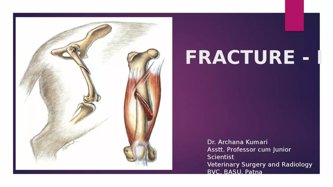
Kumari Asstt Professor cum Junior Scientist Veterinary Surgery and Radiology BVC BASU Patna Development of Bone Bone is a specialization connective tissue with mineralized collagenous framework providing skeletal support to the body ID: 932423
Download Presentation The PPT/PDF document "FRACTURE - I . Dr. Archana" is the property of its rightful owner. Permission is granted to download and print the materials on this web site for personal, non-commercial use only, and to display it on your personal computer provided you do not modify the materials and that you retain all copyright notices contained in the materials. By downloading content from our website, you accept the terms of this agreement.
Slide1
FRACTURE - I
.
Dr. Archana
Kumari
Asstt
. Professor cum Junior Scientist
Veterinary Surgery and Radiology
BVC, BASU, Patna
Slide2Development of Bone
Bone is a specialization connective tissue with mineralized collagenous framework providing skeletal support to the body.acts as lever for
muscles gives form to soft tissues protects the vital organs and
helps
in the locomotion of the
animals
also
serves as a mineral bank.
Embryonic stage development of a bone takes place from the mesenchyme, a diffuse loose cellular tissue lying between ectoderm& endoderm of the embryo
Slide3Composition of boneThe bone is composed of three major components
bone cells,
organic matrix and
Minerals
The bone cells include
osteoblasts,
osteocytes
osteoclasts,
These cells are derived
from undifferentiated mesenchymal cells (osteoprogenitor cells).
Slide4Mesenchymal cells also differentiate into:
Cartilage
Fascia
Ligament
Tendon
Slide5Cont.An early stage of development, the skeletal structure appear as dense concentration of mesenchymal cells that tend to take shape of particular bones
These localized areas of bone formation are called Ossification Centres
Ossification may be heteroplastic endochondral or intramembranous depending on the environment in which the bone formation takes place.A bone formed in tissue other than the skeleton is called a heteroplasic bone (exception : os penis
of dog ,
os cordis
of bovine &
os phrenic
of camel)
Slide6Types of bone
Based on the shape, bones can be classified as
long bonesshort bonesflat bonesLong bones are present in
limbs
eg
. Femur, humerus, tibia and radius etc
.
Short
bones are mainly seen in the joints-carpals and tarsals.
flat
bones are present in skull, pelvis and ribs.
Slide7Types of bones
Based on the structure, bones can be classified as cortical
bone compact bone
cancellous or spongy
bone
Compact bone is found largely in the shafts or long bones that surround the marrow cavity
.
Spongy bone is found in the vertebrae, flat bones and in the ends of long bones.
Slide8Intramembranous Ossification
Mesenchymal cells differentiate into osteoblastsOsteoblasts begin to secrete intercellular substance of the bone intercellular
substance replaces amorphous fluid and encloses the osteoblasts and are called as osteocytes Actively secreting osteoblasts, line the surface of newly formed spicules of the bone and continue to grow in a radial manner producing trabeculaeBranching and forming of different trabeculae result in scaffolding
The bone formed by the scaffolding trabeculae is termed as
cancellous bone
The continued deposition of fresh bone lamellae on trabeculae & appositional bone growth forms the
compact bone
T
he surface connective tissue becomes the
periosteum
Slide9Endochondral Ossification
The process by which the bone is formed in a cartilaginous environment is called endochondral ossificationMesenchymal cells differentiate into
chondroblastsCartilage model increases in length and width by interstitial as well as appositional growthIt involves :-
proliferation
Maturation
Enlargement of chondrocytes
Slide10Cont.The earliest chondrocytes of the centre matures,
Enlarge and secrete alkaline phosphatase into the intercellular substance and becomes calcifiedA new vascular environment is created which with the invasion of perichondrium by blood vessels
it brings change in the behaviour of pluripotential cells of perichondriumThese cells differentiate into osteoblasts and a thin layer of bone is laid around the cartilage .Membrane enclosing the cartilage model is called periosteum.
Slide11Cont.
formation of secondary ossification centre helps in the growth of boneThe young cartilage at each end of the model continues to grow and extend by interstitial growth to increase the length of the model
The cancellous bone in the central part is reabsorbed to form a medullary cavity and later on filled with myeloid tissue.
Slide12Anatomy Of Long BoneLong bone is divided into three parts :
Epiphysis : present on both the ends of a bone and is composed of trabeculae
Metaphysis : composed of trabecular bone which contains haematopoietic elements
Diaphysis :
hollow tube of cortical (compact) bone .its central cavity contains the medullary arterial supply
Slide131
Slide14.The entire surface of a long bone , except at the ends at which articular cartilage is present is covered by the
periosteum.The periosteum consist of an :
Outter fibrous layerInner osteogenic layer (cambium )
Slide15Blood supply
Divided into 3 main compartments:-Afferent vascular system
– arteries and arterioles carrying nutrientsEfferent vascular
system
– veins and
veinules
carrying waste products
Intermediate
vascular
system
– capillaries linking afferent and efferent
vascular system