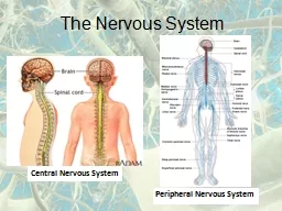
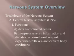
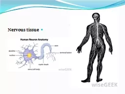
SYSTEM By Shifaa Al Qaqa Infectious agents may reach the nervous system through several routes of entry Hematogenous spread mc Direct implantation Local extension Peripheral ID: 931923
Download Presentation The PPT/PDF document "INFECTIONS OF THE NERVOUS" is the property of its rightful owner. Permission is granted to download and print the materials on this web site for personal, non-commercial use only, and to display it on your personal computer provided you do not modify the materials and that you retain all copyright notices contained in the materials. By downloading content from our website, you accept the terms of this agreement.
Slide1
INFECTIONS OF THE NERVOUSSYSTEM
By:
Shifaa
’ Al
Qa’qa
’
Slide2Infectious agents may reach the nervous system through several routes of entry:
Hematogenous
spread---- mc
Direct implantation
Local extension
Peripheral
nerves----
rabies, herpes
zoster viruses
Slide3Meningitis
Meningitis
is an inflammatory process involving the
leptomeninges
Meningoencephalitis
: infection spreads into the underlying brain
Slide4Slide5Infectious meningitis
:
- acute pyogenic (usually bacterial),
- aseptic (usually viral),
Chronic (usually
tuberculous
,
spirochetal
, or
cryptococcal
)
Chemical meningitis
Slide6Acute Pyogenic Meningitis (Bacterial Meningitis)
Neonates
: - Escherichia coli
- group B streptococci
Adolescents/young adults
: Neisseria
meningitidis
Older individuals
: - Streptococcus
pneumoniae
- Listeria
monocytogenes
Meningeal irritation
Slide7Slide8Lumbar puncture reveals an increased pressure;
examination of the CSF shows abundant neutrophils, elevated protein, and reduced glucose. Bacteria may be seen on a smear or can be cultured
Slide9Slide10MORPHOLOGY:
exudate
is evident within the
leptomeninges
over the surface of the brain
focal
cerebritis
Ventriculitis
Abscesses
Phlebitis, venous occlusion and hemorrhagic infarction
Slide11Slide12Untreated pyogenic meningitis is often fatal, but with prompt diagnosis and administration of appropriate
antibiotics
, many patients can be saved.
Slide13Aseptic Meningitis (Viral Meningitis)
The clinical course is less fulminant than in pyogenic meningitis
examination of the CSF often shows lymphocytosis, moderate protein elevation, and a normal glucose level.
self-limiting
Slide14There are no distinctive macroscopic characteristics except for brain swelling, seen in only some instances.
On microscopic examination, there is either no recognizable abnormality or a mild to moderate
leptomeningeal
lymphocytic infiltrate.
Slide15Chronic Meningitis
Tuberculous
Spirochetal
Cryptococcal
(fungus)
involve the brain parenchyma
Slide16Tuberculous Meningitis
There is only a moderate increase in CSF cellularity,
with mononuclear cells or a mixture of
polymorphonuclear
and mononuclear cells;
the protein level is elevated, often strikingly so,
and the glucose content typically is moderately reduced or normal
Slide17Infection with Mycobacterium tuberculosis also may result in a
wellcircumscribed
intraparenchymal
mass (
tuberculoma
), which may be associated with meningitis.
Chronic
tuberculous
meningitis is a cause of arachnoid fibrosis, which may produce
hydrocephalus
.
Slide18MORPHOLOGY:
The subarachnoid space contains a gelatinous or
fibrinous
exudate
, most often at the base of the brain.
Discrete white granules scattered over the
leptomeninges
.
Obliterative
endarteritis.
lymphocytes, plasma cells, and macrophages.
Florid cases show well-formed granulomas, often with
caseous
necrosis and giant cells, similar to the lesions of
tuberculosis elsewhere.
Slide19Slide20Spirochetal Infections
Neurosyphilis
Neuroborreliosis
Slide21Neurosyphilis
:
- a tertiary stage of syphilis,
- occurs in about 10% of persons with untreated
Treponema
pallidum
infection.
Slide22The infection can produce:
chronic meningitis
(
meningovascular
neurosyphilis
):
- usually involving the base of the brain,
- often with an
obliterative
endarteritis rich in plasma cells and lymphocytes.
Slide23Parenchymal involvement by spirochetes (
paretic
neurosyphilis
):
leading to neuronal loss and marked proliferation of rod shaped microglial cells.
progressive loss of mental and physical functions,
mood alterations (including delusions of grandeur),
eventually severe dementia
Slide24Tabes
dorsalis
,
resulting from damage to the
sensory nerves
in the dorsal roots,
produces impaired joint position sense and ataxia (
locomotor
ataxia
);
loss of pain sensation, leading to skin and joint damage (
Charcot joints
);
other sensory disturbances, particularly characteristic “
lightning pains
”;
and the absence of deep tendon reflexes.
Slide25Neuroborreliosis
:
- involvement of the nervous system by the spirochete
Borrelia
burgdorferi
--- Lyme disease
- Neurologic signs and symptoms are highly variable and include:
aseptic meningitis,
facial nerve palsies,
mild encephalopathy,
polyneuropathies
Slide26Slide27Slide28Parenchymal
Infections
Slide29viral
infections ----- diffuse
Bacterial infections ----- localized
other organisms ----- mixed
patterns.
In immunosuppressed
hosts, more widespread
involvement with
any agent is typical.
Slide30Brain Abscesses
Bacterial infections
These can arise
by:
- direct
implantation of organisms,
- local
extension from adjacent foci (
mastoiditis
) -
hematogenous
spread ---- acute bacterial
Endocarditi
(
septic emboli), bronchiectasis, cyanotic congenital
heart disease
Slide31progressive focal deficits
increased
intracranial
pressure
herniation
CSF white cell count and protein levels are usually
high, while
the glucose content tends to be
normal
Abscess rupture
can lead to
ventriculitis
, meningitis, and
venous sinus
thrombosis
Slide32MORPHOLOGY:
Abscesses are discrete lesions with central
liquefactive
necrosis
and a surrounding fibrous
capsule.
On
microscopic examination, the necrotic center is
surrounded by
edema and granulation tissue, often with
exuberant vascularization
. Outside the fibrous capsule is a zone
of reactive
gliosis.
Slide33Slide34Viral Encephalitis
better termed
meningoencephalitis
(
almost invariably
associated with
meningeal inflammation)
Encephalitis:
Patients
develop
generalized neurologic
symptoms, such as seizures,
confusion, delirium
, and stupor or coma, as well as focal signs,
such as
reflex asymmetry and ocular palsies
Slide35The
most characteristic histologic
features are:
- perivascular and parenchymal mononuclear cell infiltrates
- microglial nodules
neuronophagia
Slide36Slide37Slide38In addition to direct infection of the nervous system, the CNS also
can be
injured
by immune
mechanisms after systemic
viral infections
.
Slide39Arboviruses
:
arthropod-borne viruses
epidemic encephalitis ----
tropical regions
Eastern and Western equine encephalitis
West
Nile virus
infection
capable of causing
serious morbidity
and high mortality
.
In severe
cases there
may be a necrotizing
vasculitis
with associated
focal hemorrhages
.
CSF????
Symptoms???
Slide40Herpesviruses
:
HSV-1
HSV-2
Varicella-zoster virus (VZV)
Slide41HSV-1
encephalitis:
-
may occur in any age group but is most common in children and young adults
- It
typically manifests with alterations in mood, memory, and behavior, reflecting involvement of the
frontal and temporal lobes
.
- MORPHOLOGY
:
- starts in, and most severely involves, the inferior and medial regions of the temporal lobes and the orbital
gyri
of the frontal lobes.
- The infection is necrotizing and often
hemorrhagic
in the most severely affected regions.
- large
eosinophilic
intranuclear
viral inclusions
(
Cowdry
type A bodies) can be found in both neurons and glial cells.
Slide42Slide43HSV-2:
- also
affects the nervous system, usually in
the form
of meningitis in adults.
- Disseminated
severe
encephalitis occurs
in many neonates born by vaginal delivery
to women
with active primary HSV genital infections.
Slide44Varicella-zoster virus (VZV
):
Causes chickenpox during primary
infection,
usually without
any evidence of
neurologic involvement.
The virus establishes latent infection
in neurons
of
dorsal root
ganglia
Reactivation in adults
manifests as
a painful, vesicular skin eruption in the
distribution of
one or a few dermatomes (
shingles
).
This usually is
a self-limited
process, but there may be a persistent
pain syndrome
in the affected region (
postherpetic
neuralgia
).
In immunosuppressed patients,
acute herpes
zoster encephalitis can occur. Inclusion bodies
can be
found in glial cells and neurons.
Slide45Cytomegalovirus
:
CMV
infects the nervous system in fetuses and
immunosuppressed persons
Intrauterine infection causes
periventricular necrosis
, followed later by
microcephaly
with
periventricular calcification
.
Adults:
CMV
produces a
subacute
encephalitis, again often most severe in the
periventricular
region.
Lesions can be hemorrhagic and
contain typical
viral inclusion
–bearing cells---
neurons, glial
cells,
ependyma
, and endothelium
Slide46Slide47Poliovirus
:
Enterovirus
---subclinical or mild gastroenteritis
in a small fraction of
cases, it
secondarily invades the nervous system and
damages
motor
neurons
in the spinal cord and brain stem (
paralytic poliomyelitis)
loss of motor
neurons-----flaccid paralysis, muscle
wasting and
hyporeflexia
in the
corresponding region of the body
Slide48In the acute
disease, death
can occur from paralysis of respiratory muscles
.
Long after the infection has resolved, typically 25 to
35 years
after the initial illness, a
postpolio
syndrome
of
progressive weakness
associated with decreased muscle bulk
and pain
can appear. The cause of this syndrome is unclear.
Slide49Slide50Slide51Rabies
Virus
:
Rabies
is a severe encephalitic infection transmitted
to humans
from rabid
animals (dogs, bats),
usually by a bite
.
Virus enters the CNS by ascending along the
peripheral nerves
from the wound site, so the incubation
period depends
on the distance between the wound and the brain. usually taking a few
months.
Slide52The disease manifests
initially with
nonspecific symptoms of malaise, headache,
and fever.
As the
infection advances, the patient shows
extraordinary CNS
excitability
;
- the
slightest touch is
painful
violent
motor responses progressing to convulsions.
Contracture of
the pharyngeal musculature may create an
aversion to
swallowing even water (hydrophobia).
Periods of mania
and stupor progress to coma and eventually
death, typically
from
respiratory failure
.
Slide53Slide54Human Immunodeficiency
Virus
:
direct
effects of virus on the nervous
system,
opportunistic infections
,
primary
CNS lymphoma
Slide55- Aseptic meningitis
HIV
encephalitis (HIVE
)
HIV-associated neurocognitive disorder (HAND
):
cognitive dysfunction ranging from mild to
fullblown
dementia.
stem from HIV infection of
microglial cells
in
the brain. This leads to activation of innate
immune
responses.
neuronal injury likely stems
from a
combination of cytokine-induced inflammation and
toxic effects
of HIV-derived proteins.
Slide56multinucleate giant cell
microglial
nodules
, HIV
Slide57Polyomavirus
:
- Progressive
multifocal
leukoencephalopathy
(PML
)
JC
virus infects
oligodendrocytes
---
demyelination
Most people
show serologic
evidence of exposure to JC virus during
childhood---- PML
results from
virus reactivation
, as the disease is restricted to
immunosuppressed persons
Slide58The lesions are patchy, irregular, ill-defined areas of
white matter
destruction
that enlarge as the disease
progresses
Each lesion is an area of
demyelination
in
the center--- lipid-laden
macrophages and
a reduced
number of axons.
At
the
edges--- enlarged
oligodendrocyte
nuclei with glassy-appearing
amphophilic
viral inclusions
.
The virus
also infects astrocytes, leading to bizarre giant forms
with irregular,
hyperchromatic
, sometimes multiple
nuclei.
Slide59Slide60Patients develop focal and
progressive
neurologic symptoms and
signs,
Imaging studies
show extensive, often multifocal,
ring-enhancing lesions
in the hemispheric or cerebellar white matter.
Slide61Slide62Fungal Encephalitis
Candida
albicans
:
- multiple
microabscesses
,
- with
or without granuloma formation
Mucormycosis
:
infection
of
the nasal
cavity or sinuses of a diabetic
patient.
spread to the brain through vascular
invasion or
by
direct
extension through the cribriform
plate.
Aspergillus
fumigatus
:
widespread septic hemorrhagic infarctions because
of its marked
predilection for blood vessel wall
invasion and
subsequent thrombosis
Cryptococcus
neoformans
: soap bubble”–like appearance,
mucoid
encapsulated yeasts
Slide63Slide64Other Meningoencephalitides
Cerebral
Toxoplasmosis:
Protozoan Toxoplasma
gondii
Immunosuppressed
Slide65Newborns (triad of
chorioretinitis
, hydrocephalus, and intracranial calcifications),
the
CNS abnormalities are most severe when the infection occurs early in gestation during critical stages of brain development.
Necrosis
of periventricular lesions gives rise to
secondary calcifications
as well as inflammation and gliosis,
which can
lead to obstruction of the aqueduct of
Sylvius
and hydrocephalus
.
Slide66MORPHOLOGY:
Abscesses (multiple, cerebral
cortex)
Edema around
lesions
(ring
enhancing
lesions on CT and MRI)
free
tachyzoites
and encysted
bradyzoites
may be found at the periphery of the necrotic foci
Slide67Slide68other
Cysticercosis
Amebiasis
Slide69Prion Diseases
the agent in prion diseases is
an abnormal
form of a cellular
protein, termed
prion
protein (
PrP
).
PrPc
(normal)----
PrPsc
(
Abnormal)--- protease resistant/resistant to
proteolysis
Accumulation of
PrPsc
in neural tissue seems to be the cause of cell injury
Slide70Slide71sporadic, familial, iatrogenic, and
variant forms
of Creutzfeldt-Jakob disease (
CJD)
scrapie
in
sheep
bovine spongiform encephalopathy
in cattle (“mad cow disease”
Slide72Creutzfeldt-Jakob
Disease
:
rapidly progressive dementing illness
Sporadic in
approximately 85% of
cases.
Commonly affecting
persons older than 70 years of age, familial
forms caused
by mutations in PRNP may present in
younger people
infectious nature of
PrPsc
Slide73Slide74Slide75Slide76MORPHOLOGY:
The progression to death in CJD usually is so rapid that
there is
little, if any, macroscopic evidence of brain atrophy.
On microscopic
examination, the pathognomonic finding is
a
spongiform
transformation of the cerebral
cortex and
deep gray matter structures----- microscopic vacuoles of
varying sizes
within the
neuropil
and sometimes in the
perikaryon
of neurons-----neuronal
loss, No inflammation
Slide77Slide78Variant Creutzfeldt-Jakob Disease
Young adults
,
behavioral
disorders figured prominently in
early disease
stages
,
the neurologic syndrome
progressed more
slowly than in other forms of
CJD
is a consequence of
exposure to
the prion disease of cattle, called bovine
spongiform encephalopathy????
+
cortical amyloid
plaques
Slide79