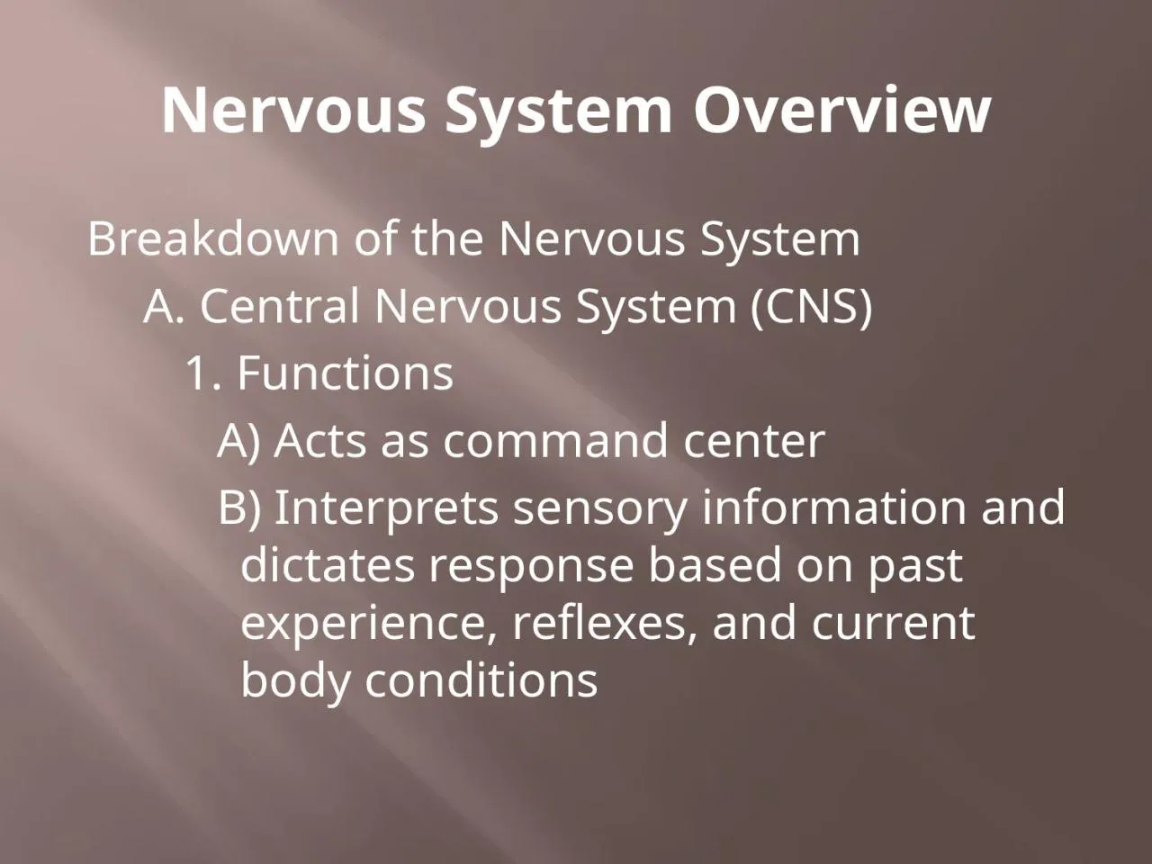
/
Nervous System Overview Breakdown of the Nervous System Nervous System Overview Breakdown of the Nervous System

Nervous System Overview Breakdown of the Nervous System - PowerPoint Presentation

gelbero
gelbero . @gelbero
Follow
358 views
Uploaded On 2022-06-01

Nervous System Overview Breakdown of the Nervous System - PPT Presentation

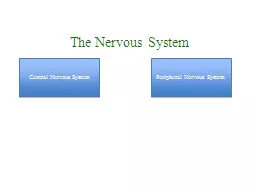
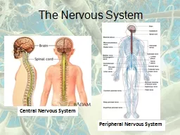
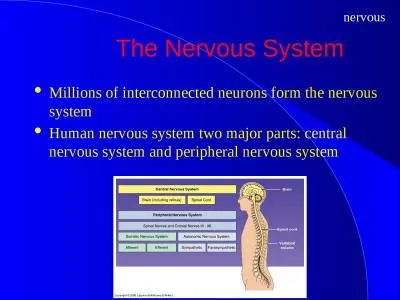
A Central Nervous System CNS 1 Functions A Acts as command center B Interprets sensory information and dictates response based on past experience reflexes and current body conditions ID: 913112

nervous system amp central system nervous central amp brain cortex sensory areas motor neurons lobes area cns anterior hemisphere

Share:

Link:

Embed:

Download Presentation from below link

Download Presentation The PPT/PDF document "Nervous System Overview Breakdown of the..." is the property of its rightful owner. Permission is granted to download and print the materials on this web site for personal, non-commercial use only, and to display it on your personal computer provided you do not modify the materials and that you retain all copyright notices contained in the materials. By downloading content from our website, you accept the terms of this agreement.


Presentation Transcript

Slide1

Nervous System Overview

Breakdown of the Nervous System

A. Central Nervous System (CNS)

1.

Functions

A)

Acts

as command center

B)

Interprets

sensory information and dictates response based on past experience,

reflexes,

and current body conditions

Slide2

Nervous System Overview

B. Peripheral Nervous System (PNS)

1.

Functions

A)

Conveys

information to and from CNS

2.

Components

A)

Somatic

sensory (afferent) neurons

1)

Carry

impulses from receptors in skin, skeletal

muscle,

and

joints to the CNS

Slide3

Nervous System Overview

B)

Visceral

sensory neurons

1)

Carry

impulses from

receptors within the visceral

organs

to the CNS

C)

Somatic

motor (efferent) neurons

1)

Carry

impulses

from the CNS to

skeletal

muscles

D)

Visceral

motor neurons

1)

Carry

impulses

from the CNS to

smooth muscle, cardiac

muscle,

and glands

Slide4

Nervous System Overview

C. D

ivisions

of PNS

1.

Somatic

NS

A)

Consists

of somatic sensory neurons and somatic motor neurons

B)

Voluntary

nervous system

2.

Autonomic

NS

A)

Consists

of visceral sensory neurons and visceral motor neurons

Slide5

Nervous System Overview

B)

Involuntary

nervous system

C) 2

branches

1) Sympathetic – fight-or-flight system

a

)

Stimulates

most effectors

2)

Parasympathetic – energy restoration/conservation system

a)

Inhibits

most effectors

Slide6

Central Nervous System

The Central Nervous System

A. Structures of the CNS

1. Cerebrum

A)

Divided

into 2 hemispheres

1)

Each

consists of

gyri

(elevated areas), sulci (shallow depressions) and fissures

Slide7

Central Nervous System

B) 5 lobes

1)

Frontal

, parietal, occipital,

temporal,

and insula

C)

Important

structures

1)

Longitudinal

fissure (right & left hemispheres)

2)

Transverse

fissure (cerebrum & cerebellum)

3)

Central

sulcus (frontal & parietal lobes)

Slide8

Central Nervous System

a)

Precentral

gyrus (within frontal lobe)

b)

P

ostcentral

gyrus (within parietal lobe)

4)

P

arieto

-occipital sulcus (parietal &

occiptal

lobes)

5)

Lateral

sulcus (temporal & frontal/parietal lobes)

Slide9

Central Nervous System

D)

Cerebral

cortex – "conscious mind"

1)

Composed

of gray matter

2)

Involved

with memory, reasoning, intelligence, etc...

3)

C

ontrilateral

Slide10

Central Nervous System

4) Exhibits hemisphere dominance

a) Left hemisphere – most functions; 90% of population

b) Right hemisphere – artistic & musical qualities; left-handed

Slide11

Central Nervous System

5) 3 main functional areas

a)

Motor

areas

i

)

Primary

motor cortex

(a)

Found

in precentral gyrus

(b)

Responsible

for conscious movement of skeletal muscles

Slide12

Central Nervous System

ii)

Premotor

cortex

(a)

Lies

anterior to primary motor cortex

(b)

Responsible

for learned motor skills that are repeated or patterned (ex. typing)

iii)

Broca’s

area

(a)

Lies

anterior & inferior to premotor cortex

(b)

Involved

in speech production

(c)

Only

in one hemisphere (usually left)

Slide13

Central Nervous System

iv)

Frontal

eye field

(a)

Lies

anterior to premotor cortex and superior to

Broca’s

area

(b)

Responsible

for voluntary eye movements

Slide14

Slide15

Central Nervous System

b)

Sensory

areas

i

)

Primary

somatosensory cortex

(a)

Lies

in

postcentral

gyrus

(b)

Allows

for spatial discrimination

ii)

Somatosensory

association cortex

(a)

Lies

posterior to primary somatosensory cortex

Slide16

Central Nervous System

(b)

Integrates

and analyzes somatic sensory inputs (i.e. pain, touch, temp, etc.) to produce an understanding of what is being felt

iii)

Visual

area

(a)

Located

within occipital lobes

iv)

Auditory

area

(a)

Found

in temporal lobes

Slide17

Central Nervous System

v)

Olfactory

area

(a)

Found

in temporal lobes in regions known as the

uncus

vi)

Gustatory

area

(a)

Found

in parietal lobe

Slide18

Slide19

Central Nervous System

c)

Association

areas

i

)

Prefrontal

cortex

(a)

Found

in anterior portions of frontal lobe

(b)

Involved

with intellect, complex

learning,

and

personality

Slide20

Central Nervous System

ii) Gnostic area

(a) Found in undefined areas of parietal, temporal, and occipital lobes

(b) Only one per hemisphere

(c) Receives input from all sensory association areas

(d) Sends input to prefrontal cortex which adds emotions

Slide21

Central Nervous System

iii

)

Language

areas

(a)

Surround

lateral sulcus in left hemisphere

(b) 4 defined

areas

(

i

)

Wernick’s

area

(a) Associated with sounding out

unfamaliar

words

Slide22

Central Nervous System

(

ii) Broca’s area

(a)

Associated

with speech production

(iii)

Lateral

prefrontal cortex

(a)

Associated

with language comprehension and word

analysis

(iv) Lateral & ventral temporal lobes

(a) Coordinates auditory & visual aspects of language

Slide23

Central Nervous System

E

)

Cerebral

white matter

1)

Lies

deep to cortex

2)

Responsible

for communication between cortical areas and also between the cortex and lower CNS centers

3) 3 types

a)

Commissures

– connect

the right

&

left sides of

the cerebrum

Slide24

Central Nervous System

b)

Association

fibers – transmit within

the same

hemisphere

c)

Projection

fibers – run to and from lower brain areas

F) B

asal

nuclei

1)

Bundles

of subcortical gray matter deep within white matter

2)

Control

large automatic skeletal muscle contractions and produce dopamine

Slide25

Central Nervous System

2. Diencephalon – central core of brain; covered by cerebrum; 3 paired structures

A)

Thalamus

– connected by

massa

intermedia

1)

Relay

station for sensory impulses from the body

2)

All sensory information

going to somatosensory cortex

goes

through it

B)

Hypothalamus

1)

Regulates

visceral information from the body

Slide26

Central Nervous System

2)

Functions

a)

Autonomic

nervous system regulator

b)

Endocrine

system regulator

c) B

ody temperature

regulation

d)

Hunger

& thirst centers

e)

Regulates wake

-sleep cycles

f)

Emotional

response center

Slide27

Central Nervous System

3)

Walls

meet and extend to form

infundibulum

a)

Suspends

the pituitary gland

C)

Epithalamus

1)

Posterior to the thalamus

2)

Contains

pineal gland

a) S

ecretes

melatonin – regulator of wake-sleep cycles

Slide28

Central Nervous System

3. Brain Stem

A)

Midbrain

1)

Contains

cerebral

aquaduct

2)

Location

of corpora

quadragemina

a)

Causes

head movements due to sound

B)

Pons – "bridge"

1) Connection between medulla oblongata & midbrain

2

)

Contains

portions of respiratory center

Slide29

Central Nervous System

C)

Medulla

oblongata

1)

Connects the brain stem to the

spinal cord

2) Contains

cardiovascular center

3

) Contains

portions of respiratory center

4

) Brain

center responsible for hiccupping, vomiting, swallowing,

coughing,

and sneezing

Slide30

Central Nervous System

4. Cerebellum

A) 1/8 of total brain

B)

I

psilateral

C) 2 hemispheres connected by the vermis

1) 3 lobes each

a) Anterior, Posterior, and

Flocculonodular

(hidden by the posterior)

Slide31

Central Nervous System

D)

Attached

to brain stem by cerebellar peduncles

E)

Coordinates learned skeletal

muscle activities

1)

Posture

, equilibrium, learned motor skills & speech

5. Limbic System

A)

Not

an isolated part of the brain; structures span large areas around the medial aspects of the cerebral hemispheres

Slide32

Central Nervous System

B)

Our

emotional brain

1)

Amygdala

– recognizes fearful facial expressions, assesses danger and elicits fear responses

2)

Cingulated

gyrus

– plays a role in expressing our emotions through gestures and in resolving mental conflicts when frustrated

3)

Hippocampus

– plays a role in memory

Slide33

Central Nervous System

6. Ventricles of the Brain

A)

Hollow

, fluid-filled chambers of the brain

B)

Filled with cerebrospinal

fluid (CSF)

C)

Lined

with ependymal cells

D)

Contain the choroid

plexus

1) Produce/recycle and help to circulate CSF

2) Composed

primarily of ependymal

cells

Slide34

Central Nervous System

E) 4 ventricles

1) Lateral ventricles – one per cerebral hemisphere

a) S

eparated

by septum

pellucidum

2) 3

rd

ventricle – in diencephalon

a)

Connected

to lateral ventricles by

interventricular

foramen

Slide35

Central Nervous System

3) 4

th

ventricle – behind pons

a)

Connected

to 3

rd

ventricle by cerebral aqueduct

b)

Connected

to the central canal of the spinal cord

Slide36

Slide37

Central Nervous System

B. Protection of

the Brain & Spinal Cord

1.

Protected

by bone (

skull & vertebrae)

, membrane (meninges) & fluid (CSF)

2.

Meningies

– 3 CT membranes

A) Dura Mater

1)

Outermost

& strongest

2)

Attached

to the skull and vertebrae

Slide38

Central Nervous System

B) Arachnoid Mater

1)

Middle

layer

2)

Separated

from dura mater by subdural space

3)

Separated

from pia mater by subarachnoid

space

Slide39

Central Nervous System

C) Pia Mater

1) Innermost & thinnest

2) Only layer that clings to brain

Slide40

Slide41

Central Nervous System

D) Cerebrospinal

Fluid (CSF)

1)

In

& around brain and

spinal cord

2)

Produced

by the choroid plexus (ependymal cells)

3)

Derived from, and similar to,

blood plasma

but

with

fewer proteins and different ion concentrations

Slide42

Central Nervous System

4)

Cushions

the brain

5)

Helps

nourish brain and eliminate waste products

Slide43

Slide44

Central Nervous System

C. Spinal Cord

1. External Anatomy

A) 2 enlargements corresponding with their location

1)

Cervical

enlargement

a)

Nerves

to and from upper limbs leave and enter here

Slide45

Central Nervous System

2)

Lumbar

enlargement

a)

Nerves

to and from lower limbs leave and enter here

B)

C

onus

medularis

1)

Cone

-like enlargement of the spinal cord inferior to the

lumbar

enlargement

C)

C

auda

equina

1)

Numerous

nerves extending inferiorly from the

conus

medularis

Slide46

Central Nervous System

D)

Spinal

nerves are connected to the SC via two bundles of axons known as roots

1)

Dorsal

root – contains the axons of sensory neurons

a)

Dorsal

root ganglion – bundle of sensory cell bodies located within the dorsal root

Slide47

Central Nervous System

2)

Ventral

root – contains the axons of motor neurons

2. Internal Anatomy

A)

Anterior

median fissure

B)

Posterior

median sulcus

C)

Gray

commissure

1)

Central

canal

D)

White commissures

Slide48

Central Nervous System

E)

Horns

1)

Anterior

,

posterior,

and lateral

F)

Columns

1)

Anterior

,

posterior,

and lateral

3. Physiology

A)

Functions

1)

Transmits impulses to and from the brain

2)

Integration

center for reflexes