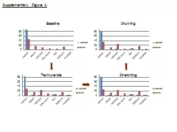
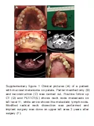
PPT-A B C D E F Supplementary figure 1 Clinical pictures (A) of a patient with mucosal melanoma
Author : Sunshine | Published Date : 2022-07-28
maxillectomy B and reconstruction C was carried out Routine follow up CT D and PETCTE shows neck node metastasis on left level Ⅳ white arrow shows the metastatic
Presentation Embed Code
Download Presentation
Download Presentation The PPT/PDF document "A B C D E F Supplementary figure 1 Clini..." is the property of its rightful owner. Permission is granted to download and print the materials on this website for personal, non-commercial use only, and to display it on your personal computer provided you do not modify the materials and that you retain all copyright notices contained in the materials. By downloading content from our website, you accept the terms of this agreement.
A B C D E F Supplementary figure 1 Clinical pictures (A) of a patient with mucosal melanoma: Transcript
maxillectomy B and reconstruction C was carried out Routine follow up CT D and PETCTE shows neck node metastasis on left level Ⅳ white arrow shows the metastatic lymph node Modified radical neck dissection was performed and implant surgery was done on upper left area 3 years after surgery F . Supplementary Figure 2.YFP is appropriately expressed in the Sox10-expressing cells and their derivatives in Sox10::iCreERtransgenics. (A-B) YFP immunostaining of E12.5 pression was detected in DRG, n Supplementary Figure . 2A:. . T-wave morphology changes upon stand-up test in LQTS . patients according to genotype. . Leads. – II, III, aVF. . Supplementary Figure . 2B:. . T-wave morphology changes upon stand-up test in LQTS patients according to . Andrew Oates. Lower Limb Specialist Physiotherapist. CDDFT. Aims. Introduce the University course structure including assessment criteria.. Review the expectations within practice.. Briefly review the ‘storey so far’ of non-medical prescribing.. . . Somatic mutation spectrum. # Substitutions. # Substitutions per Mb. b. c. a. # Substitutions per Mb. Repeats. Pseudogenes. Whole genome. Splice sites. Non-coding RNA. Intron. Promoter. 5’ UTR. 3’ UTR. Instructions for using this slide set. White slides are relevant to all anatomical sites but duplicated in each section. . Coloured slides are specific to one anatomical site. Blue = anorectal. Lavender = vulvo-vaginal. PCR amplification of . Sub1 . QTL controlling submergence tolerance using Sub1-A203 marker in . BC1F1 progenies . L. Positive parent. Negative parent. MSS-8. MSS-9. MSS-28. MSS-31. MSS-37. MSS-52. MSS-76. 2 FIGURE 1 Clinical presentation of sub-nail ALM 3 Patients Hand FootSub-nailLeftRightPalmDorsumLeftRightPlantDorsum19212835363739TotalTABLE 1 Sites of acral lentiginous melanomas in the s Achievement of a high level of survival from melanoma largely depends on early diagnosis by the primary care clinician. Early diagnosis requires careful observation of the body skin surface, examinati Update 2022. melanomafocus.org. Methodology. Open recruitment of Guideline Development Group. Clinical questions. Literature search. Evidence review. Discussion and agreement of recommendations. Drafting. E. F. A. C. D. APCMin. /+. APCMin. /+/C3aR-/-. APCMin. /+/C3aR-/-. APCMin. /+. APCMin. /+. APCMin. /+/C3aR-/-. APCMin. /+. APCMin. /+/C3aR-/-. APCMin. /+/C3aR-/-. APCMin. /+. APCMin. /+. APCMin. /+/C3aR-/-. Gabriel C, Blome S, Malogolovkin A, Parilov S, Kolbasov D, Teifke JP, et al. Characterization of African Swine Fever Virus Caucasus Isolate in European Wild Boars. Emerg Infect Dis. 2011;17(12):2342-2345. https://doi.org/10.3201/eid1712.110430. mJ. /cm. 2. was range of UV irradiation.. Supplementary data . Figure S2. Total phenolic and flavonoid contents of DW extract of . Nypa. . fruticans. . (NFD) and 100% . EtOH. extract of . Nypa. . Consort flow chart summarizing the number of patients screened, enrolled, treated and followed on the study. *one patient was re-assessed for eligibility at recurrence and re-enrolled on the study. Created with BioRender.com.. PsaM. proteins. Blue mark indicate conservation of amino acids below 90%.. Supplementary Figure 2. . (A) Phylogenetic tree of . PsaM. genes showing five clusters (mentioned as 1, 2, 3, 4, and 5). (B).
Download Document
Here is the link to download the presentation.
"A B C D E F Supplementary figure 1 Clinical pictures (A) of a patient with mucosal melanoma"The content belongs to its owner. You may download and print it for personal use, without modification, and keep all copyright notices. By downloading, you agree to these terms.
Related Documents