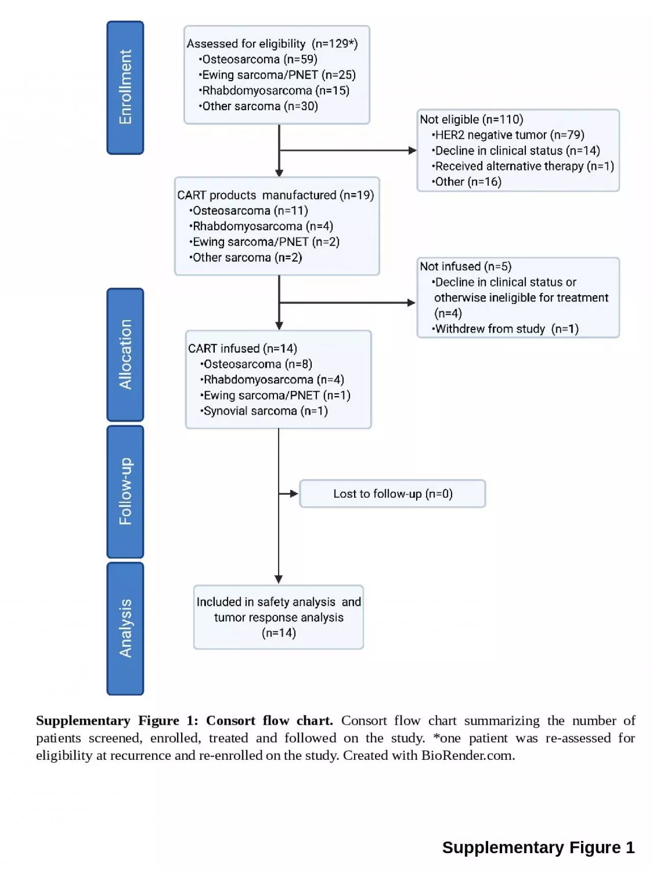
PPT-Supplementary Figure 1 Supplementary Figure 1: Consort flow chart.

Author : leah | Published Date : 2024-01-29

Consort flow chart summarizing the number of patients screened enrolled treated and followed on the study one patient was reassessed for eligibility at recurrence

Presentation Embed Code

Download Presentation

Download Presentation The PPT/PDF document "Supplementary Figure 1 Supplementary Fig..." is the property of its rightful owner. Permission is granted to download and print the materials on this website for personal, non-commercial use only, and to display it on your personal computer provided you do not modify the materials and that you retain all copyright notices contained in the materials. By downloading content from our website, you accept the terms of this agreement.

Supplementary Figure 1 Supplementary Figure 1: Consort flow chart.: Transcript


Consort flow chart summarizing the number of patients screened enrolled treated and followed on the study one patient was reassessed for eligibility at recurrence and reenrolled on the study Created with BioRendercom. Supplementary Figure 2.YFP is appropriately expressed in the Sox10-expressing cells and their derivatives in Sox10::iCreERtransgenics. (A-B) YFP immunostaining of E12.5 pression was detected in DRG, n Supplementary Figure 2: TOR downstream components ATG1 and 4EBP1 (Thor) modulate the VAP (P58S) bouton phenotype VAP (P58S) over expression using C155-Gal4 leads to larger boutons at the NMJ. (B) O Supplementary Figure S1.. Two electrodes with 1 mm gap between them were brought in contact with the surface of the water solution of 10 M NaCl in the experimental vessel. DC voltages, induced across Supplementary Figure . 2A:. . T-wave morphology changes upon stand-up test in LQTS . patients according to genotype. . Leads. – II, III, aVF. . Supplementary Figure . 2B:. . T-wave morphology changes upon stand-up test in LQTS patients according to . Figure . 1.. . A. . Gating . strategy for monocyte . identification. . PBMCs were processed as described in Figure 1 and acquired on a . FACSCalibur. flow cytometer. . Left panel:. Initial . cell gate was delineated on . washed repeatedly in PBS and PBS containing 0.3% Triton X-100 (PBS/Triton), and then incubated in blocking buffer (5% normal goat serum, 0.2% cold white fish skin gelatin, PBS/Triton) for 1 hour. Sections were then incubated with mouse anti-tyrosine hydroxylase (1:1000; Millipore; Billerica, MA) for 48 hours at 4°C. They were subsequently washed five times in PBS/Triton before being incubated with . . . Somatic mutation spectrum. # Substitutions. # Substitutions per Mb. b. c. a. # Substitutions per Mb. Repeats. Pseudogenes. Whole genome. Splice sites. Non-coding RNA. Intron. Promoter. 5’ UTR. 3’ UTR. : Crystal structure B - factor distribution and electron density quality. a) Rendering of YaxA, PaxB and YaxB crystal structures by B - factor values. For PaxB, all four NCS - related copies are show YIV-906. (500mg/kg . p.o.. bid . x7. days) plus different dose of anti-PD1 . (70ug or 200ug/mice . i.p. . once) . against . Hepa. 1-6 in vivo.. Tumor size at the beginning were about . 270mm. 3. hCMEC. /D3 cells. The TEM shows one astrocyte in direct contact with the endothelium. . Scale bar = 2. μ. m.. Endothelium. Astrocyte. Collagen gel. Supplementary figure 2. Transmission electron micrograph of . PCR amplification of . Sub1 . QTL controlling submergence tolerance using Sub1-A203 marker in . BC1F1 progenies . L. Positive parent. Negative parent. MSS-8. MSS-9. MSS-28. MSS-31. MSS-37. MSS-52. MSS-76. E. F. A. C. D. APCMin. /+. APCMin. /+/C3aR-/-. APCMin. /+/C3aR-/-. APCMin. /+. APCMin. /+. APCMin. /+/C3aR-/-. APCMin. /+. APCMin. /+/C3aR-/-. APCMin. /+/C3aR-/-. APCMin. /+. APCMin. /+. APCMin. /+/C3aR-/-. mJ. /cm. 2. was range of UV irradiation.. Supplementary data . Figure S2. Total phenolic and flavonoid contents of DW extract of . Nypa. . fruticans. . (NFD) and 100% . EtOH. extract of . Nypa. . PsaM. proteins. Blue mark indicate conservation of amino acids below 90%.. Supplementary Figure 2. . (A) Phylogenetic tree of . PsaM. genes showing five clusters (mentioned as 1, 2, 3, 4, and 5). (B).

Download Document

Here is the link to download the presentation.
"Supplementary Figure 1 Supplementary Figure 1: Consort flow chart."The content belongs to its owner. You may download and print it for personal use, without modification, and keep all copyright notices. By downloading, you agree to these terms.

Related Documents