/
The Nose and Paranasal Sinuses The Nose and Paranasal Sinuses

The Nose and Paranasal Sinuses - PowerPoint Presentation

SunshineSmiles
SunshineSmiles . @SunshineSmiles
Follow
344 views
Uploaded On 2022-08-02

The Nose and Paranasal Sinuses - PPT Presentation

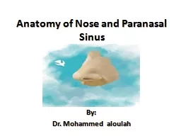
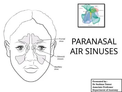
Anatomy of the Nose External Nose Pyramidal Osteocatilaginous framework covered with muscles and skin Bony Part Upper 13 2 nasal bones nasal process of frontal bone frontal processes of maxillae ID: 932654

nose nasal lateral artery nasal nose artery lateral vein cartilage posterior drains palatine anterior ethmoidal sphenopalatine bone inferior wall

Share:

Link:

Embed:

Download Presentation from below link

Download Presentation The PPT/PDF document "The Nose and Paranasal Sinuses" is the property of its rightful owner. Permission is granted to download and print the materials on this web site for personal, non-commercial use only, and to display it on your personal computer provided you do not modify the materials and that you retain all copyright notices contained in the materials. By downloading content from our website, you accept the terms of this agreement.


Presentation Transcript

Slide1

The Nose and Paranasal Sinuses

Slide2

Anatomy of the Nose

External Nose:

Pyramidal

Osteocatilaginous framework, covered with muscles and skin.Bony Part: Upper 1/32 nasal bones, nasal process of frontal bone, frontal processes of maxillae.Cartilaginous part:Upper lateral Cartilage: it’s lower free edge forms part of the nasal valve.Lower lateral cartilage (alar cartilage): U-shaped, Medial and lateral crura.Lesser alar (Sesamoid) cartilage: 2 or moreSeptal cartilage

Slide3

Anatomy of the Nose

Internal Nose:

Divided into left and right nasal cavities by the nasal septum

Each nasal cavity communicates with the exterior through the nasris or nostril, and with the nasopharynx through the posterior nasal aperture or the choana.Each nasal cavity consists of skin lined portion (the vestibule) and a mucosa lined portion (the nasal cavity proper).

Slide4

Anatomy of the Nose

Internal Nose:

Vestibule:

Anterior and inferior part of the nasal cavityLined by skin, contains sebaceous glands, hair follicles, hair (vibrissae)Nasal valveNasal Cavity Proper:Lateral nasal wall: 3 turbinates, with 3 corresponding meati (space bellow the turbinate)Inferior meatus: drains the nasolacrimal duct.Middle meatus: drains the maxillary, frontal, anterior ethmoidal sinusesSuperior meatus: drains the posterior ethmoidal sinuses.The nasal septum (medial wall): Bone: vomer, perpendicular plate of ethmoid bone, maxillary crest, palatine bone

Cartilage: quadrangular cartilageRoof: nasal bone anteriorly, cribriform plate of ethmoid in the middle, body of sphenoid posteriorly.Floor: palatine process of maxilla, palatine bone.

Slide5

Lateral Nasal Wall

Slide6

Nasal Septum

Slide7

Lining of the nose:

Vestibule: skin

Olfactory area

Respiratory region: pseudostratified ciliated columnar epithelium, erectile, highly vascular.Blood supply:Internal and external carotid arteries.Nasal septum:ICA: anterior and posterior ethmoidal arteries.ECA: sphenopalatine artery, greater palatine artery, superior labial artery.Lateral wall:ICA: anterior and posterior ethmoidal arteries.ECA: sphenopalatine artery, greater palatine artery, maxillary artery, facial artery.

Slide8

Kiesselbach’s

plexus (Little’s area)

Confluence of vessels along the anterior nasal septum where the septal branch of sphenopalatine artery, anterior ethmoidal artery branches, greater palatine artery, and septal branches of superior labial artery anastomose

Woodruff’s plexus (naso -nasopharyngeal plexus)Anastomosis of posterior ethmoid, sphenopalatine, and ascending pharyngeal arteries along posterior lateral nasal wall inferior to the inferior turbinate

Slide9

Slide10

Venous Drainage

Sphenopalatine vein drains via sphenopalatine foramen into pterygoid plexus.

Ethmoidal veins drain into superior ophthalmic vein.

Venous system is valvelessAnterior facial vein drains through common facial vein to internal jugular vein; also communicates with cavernous sinus via ophthalmic veinsAngular vein drains external nose via ophthalmic vein to cavernous sinus.Lymphatic DrainageAnterior portion of nose drains toward external nose in the submandibular nodes.Posterior portion into upper deep cervical nodes.

Slide11

Slide12

Anatomy

External nasal valve (nasal vestibule) formed by:

columella

Nasal rim (caudal border of the lower lateral cartilage). nasal floorThe nasalis muscle dilates this portion during inspiration.Internal nasal valve formed by:Nasal septumcaudal border of the Upper lateral Cartilagehead of the inferior turbinate

Slide13

Paranasal sinuses

Slide14

Paranasal sinuses

Pseudostratified columnar ciliated epithelium with goblet cells

Nasal cilia beat 10-20 times/sec at room temp

Functions; Humidification, Vocal resonance, Mucus production, Absorbs shock to the head, Regulation of intranasal pressureThe ethmoid and the maxillary sinuses; Present at birth

Slide15

Slide16

R1

Slide17

R2

Slide18

R3

Slide19

R4