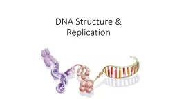
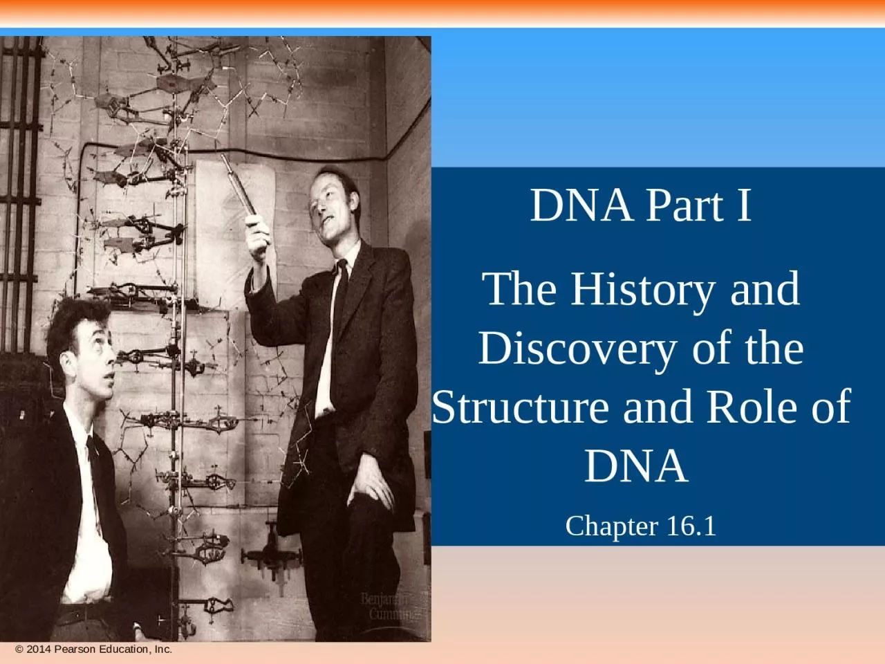
Chapter 161 Life s Operating Instructions In 1953 James Watson and Francis Crick introduced an elegant doublehelical model for the structure of deoxyribonucleic acid or DNA Hereditary information is encoded in DNA ID: 934082
Download Presentation The PPT/PDF document "DNA Part I The History and Discovery of ..." is the property of its rightful owner. Permission is granted to download and print the materials on this web site for personal, non-commercial use only, and to display it on your personal computer provided you do not modify the materials and that you retain all copyright notices contained in the materials. By downloading content from our website, you accept the terms of this agreement.
Slide1
DNA Part I
The History and Discovery of the Structure and Role of DNA Chapter 16.1
Slide2Life’s Operating InstructionsIn 1953, James Watson and Francis Crick introduced an elegant double-helical model for the structure of deoxyribonucleic acid, or DNAHereditary information is encoded in DNA and is found in the nucleus of every cell in the bodyDNA contains the instructions to build every trait in an organism. This includes biochemical, anatomical, physiological, and(to some extent) behavioral traits.
Slide3DNA is the genetic materialEarly in the 20th century, the identification of the molecules of inheritance loomed as a major challenge to biologists
Slide4Identifying DNA as a unique
molecule
1869- Friedrich
Miescher
, a
Swiss chemist
was
the first to identify DNA as a unique molecule
.
S
howed
that when
the enzyme pepsin was
used on the nucleus of
blood cells, a
strange phosphorous-containing material remained.
He
called this molecule
nuclein
, but still
thought that proteins were the molecules of heredity.
Slide5Genes are Located on chromosomesIn 1920, Thomas Hunt Morgan showed that genes are located on chromosomes. The two components of chromosomes—DNA and protein—became candidates for the genetic material
Slide6Evidence That DNA Can Transform BacteriaThe discovery of the genetic role of DNA began with research by Frederick Griffith in 1928Griffith worked with two strains of a bacterium, one pathogenic and one harmlessWhen he mixed the heat-killed pathogenic strain with living cells of the harmless strain, some living cells became pathogenicHe called this phenomenon transformation. A “transforming factor” turned the harmless strain into a pathogenic one
Slide7Can a genetic trait be transferred between different bacterial strains?https://www.youtube.com/watch?v=MsFM-hvLtsE
Slide8In 1944, Oswald Avery, Maclyn McCarty, and Colin MacLeod performed a similar experiment using a different bacteria and announced that the “transforming factor” was DNAMeaning that the instructions for being “pathogenic” was contained in its DNA and could be transferred to the harmless strainMany biologists remained skeptical, mainly because little was known about DNAMost scientists still believed that protein contained genetic material
Slide9Evidence That Viral DNA Can Program CellsMore evidence for DNA as the genetic material came from studies of viruses that infect bacteriaSuch viruses, called bacteriophages (or phages), are widely used in molecular genetics researchA virus is a nucleic acid (DNA sometimes RNA) enclosed by a protective protein coat
Slide10Structure & Replication Cycle Of T2 Bacteriophage Virus
Slide11In 1952, Alfred Hershey and Martha Chase showed that DNA is the genetic material of a phage known as T2They designed an experiment showing that only one of the two components of T2 (DNA or protein) enters an E. coli cell during infectionThey concluded that the injected DNA of the phage provides the genetic information
Slide12Animation: Hershey-Chase Experiment
Slide13Slide14Additional Evidence That DNA Is the Genetic MaterialIt was already known that DNA is a polymer of nucleotides, each consisting of a nitrogenous base, a sugar, and a phosphate groupIn 1950, Erwin Chargaff reported that DNA composition varies from one species to the nextThis evidence of diversity made DNA a more credible candidate for the genetic material
Slide15Based on the observations above,
three rules can be deduced%A =%T; %C=%GA+G=C+T=50%The percentages of the nucleotide vary for different speciesThe basis for these rules was not understood until the discovery of the double helix
Chargaff’s Rules
Slide16Using Chargaff’s Rules
If thymine makes 40% of the nucleotides in a species. What percentage of nucleotides will be cytosine?
Slide17Building a Structural Model of DNAAfter DNA was accepted as the genetic material, the challenge was to determine how its structure accounts for its role in heredityMaurice Wilkins and Rosalind Franklin were using a technique called X-ray crystallography to study molecular structureFranklin produced a picture of the DNA molecule using this technique
Slide18Figure 16.6
(a) Rosalind Franklin
(b) Franklin’s X-ray diffraction
photograph of DNA
Slide19Franklin’s crystallographic images enabled Watson to deduce that DNA was helical The X-ray images also enabled Watson to deduce the width of the helix and the spacing of the nitrogenous basesThe pattern in the photo suggested that the DNA molecule was made up of two strands, forming a double helix
Slide20Franklin had concluded that there were two outer sugar-phosphate backbones, with the nitrogenous bases paired in the molecule’s interiorWatson and Crick built models of a double helix to conform to the X-rays and chemistry of DNAWatson built a model in which the backbones were antiparallel (their subunits run in opposite directions)
Slide21At first, Watson and Crick thought the bases paired like with like (A with A, and so on), but such pairings did not result in a uniform width Instead, pairing a purine with a pyrimidine resulted in a uniform width consistent with the X-ray data
Slide22Figure 16.UN02
Purine + purine: too wide
Pyrimidine + pyrimidine: too narrow
Purine + pyrimidine: width
consistent with X-ray data
Slide23Watson and Crick reasoned that the pairing was more specific, dictated by the base structuresThey determined that adenine (A) paired only with thymine (T), and guanine (G) paired only with cytosine (C); called complimentary base paringA & G are pyrimidines; C & T are purinesThe Watson-Crick model explains Chargaff’s rules: in any organism the amount of A = T, and the amount of G = CAnd it is the sequence of nucleotides in DNA that determines an organism’s traits
Slide24Figure 16.7b
(b) Partial chemical structure
3
′
end
5
′
end
3
′
end
5
′
end
Hydrogen bond
T
A
G
C
A
T
C
G
Slide25Figure 16.7a
(a) Key features of DNA structure
0.34 nm
1 nm
3.4 nm
T
T
T
A
A
A
C
C
C
G
G
G
A
T
T
A
C
G
C
G
A
T
C
G
C
G
C
G
C
G
Slide26Figure 16.5
5′ end
Thymine (T)
Adenine (A)
Cytosine (C)
Guanine (G)
Nitrogenous
bases
Sugar–
phosphate
backbone
3
′
end
Nitrogenous
base
Sugar
(
deoxyribose
)
DNA
nucleotide
Phosphate
Slide27Staining of DNA reveals somatic cells have the same amount of DNA and twice as much as gametes.
1914-Robert Feulgen, a German chemist, found a
technique that determines the relative amount
of DNA
present in a cell.
Found
that all cells in an organism had the same amount of DNA except gametes, which had half the normal amount.