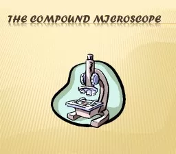
The Compound Microscope Parts of the Microscope Base bottom of microscope supports Light Source makes the specimen visible Stage Where you place the slide Arm Supports upper part of microscope ID: 774185
Download Presentation The PPT/PDF document "The Compound Microscope" is the property of its rightful owner. Permission is granted to download and print the materials on this web site for personal, non-commercial use only, and to display it on your personal computer provided you do not modify the materials and that you retain all copyright notices contained in the materials. By downloading content from our website, you accept the terms of this agreement.
The Compound Microscope
Parts of the Microscope Base – bottom of microscope, supports Light Source – makes the specimen visible Stage – Where you place the slide Arm – Supports upper part of microscope Fine Adjustment – Sharpens the image, both powers Coarse Adjustment – Focuses the image under low power Eyepiece (Ocular) – What you look through, 10x
Parts of the Microscope Body Tube – Attaches eyepiece to nosepiece (objectives) Revolving nosepiece – holds objective lenses High power objective – provides 40x magnification Low power objectives – provides 10x magnification Stage Clips – Hold the slide in place Diaphragm – Controls/regulates the amount of light
Eyepiece (Ocular) Body Tube Revolving nosepiece H.P. Objective L.P. Objective Stage Clips Diaphragm Light Source Base Stage Arm Fine Adjustment Course Adjustment
Anton van Leeuwenhoek Developed the first simple microscope One lens First to see “ Cells ”
Robert Hooke Made a compound microscope Two lenses Observed thin slices of cork Came up with the term “cells”
1. Total Magnification = Eyepiece (ocular) x Objective 10x x 25x = 250 Magnification of our microscopes Eyepiece – 10xScanning – 4xLow – 10xHigh – 40x
2. Objects appear… Larger upside down backwards “Microscope”
3. Field of View – circle of light How much of the objects you are able to see in the microscope Less of the object is seen As you go from low to high power: Field of view gets Smaller (more magnified) Darker (less light)
4. Movement of Objects Your specimen will move in the opposite direction you move the slide Due to two lenses being used If you move the slide to the left, the object will… Move to the right If you move the slide up, the object will… Move down
5. Making a Wet Mount slide Place a drop of water on the slide Place specimen on top of water Touch coverslip to water at an angle and slowly lower down prevents air bubbles
6. Adding stain or solution Do NOT remove cover slip Touch paper towel to edge of cover slip Drop solution or stain on opposite edge of cover slip
7. Estimating the size of an object Use the following equation Diameter of the Field of View # of objects that fit across
7. Estimating the size of an object ( con’t ) 2 mm ________ mm .67 2 mm 3
Using a Microscope – Rules to Follow Plug in and turn on Microscope Always start with the scanning or low power objective Place slide on the center of the stage Use Coarse Adjustment knob to focus specimen Then use Fine Adjustment to sharpen image Object now in focus
Using a Microscope – Rules to Follow When switching to a higher power objective Make sure your specimen is centered in the field of view Use tip of pointer, in your field of view, as a guide Turn nosepiece to next highest objective Focus the specimen Scanning to low use coarse adj. then fine adj.Low to High use fine adj. only