/
Spirochetes in the Context of Their Environment and Other Microbes Spirochetes in the Context of Their Environment and Other Microbes

Spirochetes in the Context of Their Environment and Other Microbes - PowerPoint Presentation

adia
adia . @adia
Follow
66 views
Uploaded On 2023-07-09

Spirochetes in the Context of Their Environment and Other Microbes - PPT Presentation

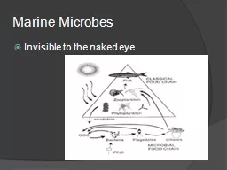
Author Michael Witty Citation Michael Witty 2009 Spirochetes in the context of their environment and other microbes Publication Date August 2009 Introduction Most observations of spirochetes come from cultivated microbes or rich animal systems  This is because of the small size of these ID: 1007276

soil spirochetes context microbes spirochetes soil microbes context microbiol 2009 treponema complex samples motion approximately environment water role large

Share:

Link:

Embed:

Download Presentation from below link

Download Presentation The PPT/PDF document "Spirochetes in the Context of Their Envi..." is the property of its rightful owner. Permission is granted to download and print the materials on this web site for personal, non-commercial use only, and to display it on your personal computer provided you do not modify the materials and that you retain all copyright notices contained in the materials. By downloading content from our website, you accept the terms of this agreement.


Presentation Transcript

1. Spirochetes in the Context of Their Environment and Other MicrobesAuthor: Michael WittyCitation: Michael Witty. 2009. Spirochetes in the context of their environment and other microbes.Publication Date : August 2009

2. IntroductionMost observations of spirochetes come from cultivated microbes or rich animal systems.  This is because of the small size of these bacteria and the technical difficulties associated with resolving them in complex environmental samples.  This movie shows bacteria from a soil sample swimming amongst soil particles and other soil microbes.Spirochetes are usually only observed after culture in vitro or from animal environments like guts (5).  However, they are widely distributed in nature and presumably play an important role as free living microbes in environments such as soil.  For example, analysis of soil communities show spirochetes contribute approximately 1% of diversity (6), a large amount for such a complex ecosystem.Spirochetes are near the limits of optical microscopy (with some dimensions of approximately 1 µm) and so are hard to see, especially when the environmental context of samples is preserved, including soil particles and large protozoa.  Bulky samples must contain many planes of foci, allowing the very small spirochetes to swim out of view with ease.  This movie shows spirochetes in the context of their fellow microbes and their natural soil environment.     Methods Water from a temporary puddle was collected using a Pasteur pipette to draw material from the surface of submerged mud, then transported to the laboratory in an Eppendorf tube.  Methyl cellulose (Protoslo; Carolina Biological Supply Company, Burlington, NC) was used to reduce the motion of microbes and increase ease of observation (1).  Approximately 50 µl of water was mixed with 1% methyl cellulose on a microscope slide and then a cover slip was applied.  The slide was examined using conventional bright-field microscopy.  When spirochetes were located, their motion was recorded using a Pupil Cam attachment (Ken-A-Vision, Kansas City, MO) and then replayed using slow motion. Discussion Spirochetes are well known as pathogens such as Treponema pallidum (syphilis) (7), Treponema pertenue (yaws) (2), Leptospira sp. (leptospirosis) (8), Borrelia burgdorferi (Lyme disease) (4), and Borrelia recurrentis (relapsing fever) (3).  However, few biologists appreciate that spirochetes are widespread in nature and play a significant role in benevolent ecological processes. References 1.  Aufderheide, K. J.  2007.  An overview of techniques for immobilizing and viewing living cells.  Micron 39(2):71–76. 2.  Backhouse, J. L., B. J. Hudson, P. A. Hamilton, and S. I. Nesteroff.  1998.  Failure of penicillin treatment of yaws on Karkar Island, Papua New Guinea.  Am. J. Trop. Med. Hyg. 59(3):388–392. 3.  Cutler, S. J., A. Abdissa, and J. F. Trape.  2009.  New concepts for the old challenge of African relapsing fever borreliosis.  Clin. Microbiol. Infect. 15 :400–406. 4.  Gasser, R., E. Reisinger, B. Eber, R. Pokan, G. Seinost, J. Berglöff, R. Horwarth, B. Sedaj, and W. Klein.  1995.  Cases of Lyme borreliosis resistant to conventional treatment: improved symptoms with cephalosporin plus specific beta-lactamase inhibition.  Microb. Drug Resist. 1(4):341–344. 5.  Harwood, C. S., and E. Canale-Parola.  1984.  Ecology of spirochetes.  Ann. Rev. Microbiol. 38:161–192. 6. Hill, J. E., R. P. Seipp, M. Betts, L. Hawkins, A. G. van Kessel, W. L. Crosby, and S. M. Hemmingsen.  Extensive profiling of a complex microbial community by high-throughput sequencing.  Appl. Environ. Microbiol. 68:3055–3066. 7.  Radolf, J. D., and D. C. Desrosiers.  2009.  Treponema pallidum, the stealth pathogen, changes, but how?  Mol. Microbiol. 72 :1081–1086. 8.  Sohan, L., B. Shyamal, T. S. Kumar, M. Malini, K. Ravi, V. Venkatesh, M. Veena, and S. Lal.  2008.  Studies on Leptospirosis outbreaks in Peddamandem Mandal of Chittoor district, Andhra Pradesh.  J. Communic. Dis. 40(2):127–132.

3.