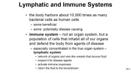
/
 21- 1 the body harbors about 10,000 times as many bacterial cells as human cells  21- 1 the body harbors about 10,000 times as many bacterial cells as human cells

21- 1 the body harbors about 10,000 times as many bacterial cells as human cells - PowerPoint Presentation

alexa-scheidler
alexa-scheidler . @alexa-scheidler
Follow
344 views
Uploaded On 2020-04-04

21- 1 the body harbors about 10,000 times as many bacterial cells as human cells - PPT Presentation

some beneficial some potentially disease causing immune system not an organ system but a population of cells that inhabit all of our organs and defend the body from agents of disease especially concentrated in the true organ system ID: 775416

lymph lymphatic cells tissue lymph lymphatic cells tissue nodes system hill companies mcgraw organs display figure fluid blood reproduction

Share:

Link:

Embed:

Download Presentation from below link

Download Presentation The PPT/PDF document " 21- 1 the body harbors about 10,000 tim..." is the property of its rightful owner. Permission is granted to download and print the materials on this web site for personal, non-commercial use only, and to display it on your personal computer provided you do not modify the materials and that you retain all copyright notices contained in the materials. By downloading content from our website, you accept the terms of this agreement.


Presentation Transcript

Slide1

21-1

the body harbors about 10,000 times as many bacterial cells as human cellssome beneficialsome potentially disease causingimmune system – not an organ system, but a population of cells that inhabit all of our organs and defend the body from agents of diseaseespecially concentrated in the true organ system – lymphatic systemnetwork of organs and vein-like vessels that recover fluidinspect it for disease agentsactivate immune responsesreturn the fluid to the bloodstream

Unit 10

Lymphatic

and Immune

Systems

Slide2

21-2

maintain fluid balanceprotect body from infection and disease

Lymphatic and Immune Systems

Figure 21.3a

(a)

Arteriole

Capillary bed

Tissue fluid

Tissue cell

Lymphatic

capillary

Venule

Copyright © The McGraw-Hill Companies, Inc. Permission required for reproduction or display.

Slide3

21-3

fluid recoveryfluid continually filters from the blood capillaries into the tissue spacesblood capillaries reabsorb 85%15% (2 – 4 L/day) of the water and about half of the plasma proteins enters lymphatic system and then returned to the bloodimmunityexcess filtered fluid picks up foreign cells and chemicals from the tissuespasses through lymph nodes where immune cells stand guard against foreign matteractivate a protective immune responselipid absorptionlacteals in small intestine absorb dietary lipids that are not absorbed by the blood capillaries

Functions of Lymphatic System

Slide4

21-4

Lymphclear, colorless fluid, similar to plasma, but much less proteinextracellular fluid drawn into lymphatic capillariesthe recovered fluidlymphatic vessels -similar to veins, have valvestransport the lymphlymphatic tissuescomposed of aggregates of lymphocytes and macrophages that populate many organs in the bodylymphatic organsdefense cells are especially concentrated in these organsseparated from surrounding organs by connective tissue capsules

Components of the Lymphatic System

Slide5

21-5

lymphatic capillaries

(terminal lymphatics)

penetrate nearly every tissue of the body

absent from central nervous system, cartilage, cornea, bone and bone marrow

sacs of thin endothelial cells that loosely overlap each other

closed at one end

cells tethered to surrounding tissue by protein filaments

gaps between cells are large enough to allow bacteria and cells entrance to lymphatic capillary

endothelium creates valve-like flaps that open when interstitial fluid pressure is high, and close when it is low

Slide6

21-6

Lymphatic Capillary

Figure 21.3b

(b)

Opening

Lymph

Tissue

fluid

Endothelium

of lymphatic

capillary

Anchoring

filaments

Copyright © The McGraw-Hill Companies, Inc. Permission required for reproduction or display.

Slide7

21-7

Route of Lymph Flow

lymphatic capillaries

collecting vessels

:

course through many lymph nodes

six lymphatic trunks

:

drain major portions of body

two collecting ducts

:

right lymphatic duct

– receives lymph from right arm, right side of head and thorax; empties into right subclavian vein

thoracic duct

- larger and longer, begins as a prominent sac in abdomen called the

cisterna chyli

; receives lymph from below diaphragm, left arm, left side of head, neck, and thorax; empties into left subclavian vein

subclavian veins

Slide8

21-

8

The Fluid Cycle

Figure 21.1

Figure 21.5

Subclavian vein

Cardiovascular system

Lymphatic system

Lymphatic

capillaries

Lymph

nodes

Lymphatic

trunks

Collecting

duct

Collecting

vessels

Lymph

flow

Lymphatic

capillaries

Pulmonary

circuit

Superior

vena cava

Blood

flow

Systemic

circuit

Axillary lymph node

Spleen

Inguinal lymph nodes

Cervical lymph nodes

R. lymphatic duct

Thymus

Thoracic duct

Cisterna chyli

Palatine tonsil

Red bone marrow

Thoracic duct

Intestinal trunk

R. and l. lumbar trunks

Popliteal lymph nodes

Abdominal,

intestinal,

and mesenteric

lymph nodes

Lymphatic vessels

L. internal jugular v.

Copyright © The McGraw-Hill Companies, Inc. Permission required for reproduction or display.

Slide9

21-9

Cells of the Lymph

natural killer (NK) cells

large lymphocytes that attack and destroy bacteria, transplanted tissue, host cells infected with viruses or have turned cancerous

responsible for immune surveillance

T lymphocytes (T cells)

mature in thymus

B lymphocytes (B cells)

activation causes proliferation and differentiation into

plasma cells

that produce

antibodies

Slide10

21-10

Lymphatic Cells

macrophages

very large, avidly phagocytic cells of the connective tissue

develop from monocytes

phagocytize tissue debris, dead neutrophils, bacteria, and other foreign matter

process foreign matter and display antigenic fragments to certain T cells alerting the immune system to the presence of the enemy

antigen presenting cells (APCs)

dendritic cells

branched, mobile APCs found in epidermis, mucous membranes, and lymphatic organs

alert immune system to pathogens that have breached their surface

reticular cells

branched stationary cells that contribute to the stroma of a lymphatic organ

act as APCs in the thymus

Slide11

21-11

Macrophages

Figure 21.7

5

µ

m

Macrophages

Pseudopods

Bacteria

Copyright © The McGraw-Hill Companies, Inc. Permission required for reproduction or display.

Peter Arnold, Inc.

Slide12

21-12

Organs of the Lymphatic System

lymphatic organs have well-defined anatomical sites

have

connective tissue capsule

that separates the lymphatic tissue from neighboring tissues

primary lymphatic organs

red bone marrow

and

thymus

site where T and B cells become

immunocompetent

– able to recognize and respond to antigens

secondary lymphatic organs

lymph nodes

,

tonsils

, and

spleen

immunocompetent cells populate these tissues

Slide13

Red Bone Marrow

red bone marrow is involved in hemopoiesis (blood formation) and immunitysoft, loosely organized, highly vascular materialseparated from osseous tissue by endosteum of boneas blood cells mature, they push their way through the reticular and endothelial cells to enter the sinus and flow away in the blood stream

21-

13

Slide14

21-14

Thymus

thymus

– member of the endocrine, lymphatic, and immune systems

houses developing lymphocytes

secretes hormones regulating their activity

bilobed organ located in superior mediastinum between the sternum and aortic arch

degeneration or involution with age

fibrous capsule gives off

trabeculae (septa)

that divide the gland into several lobes

lobes have cortex and medulla populated by T lymphocytes

reticular epithelial cells

seal off cortex from medulla forming

blood-thymus barrier

produce signaling molecules

thymosin

,

thymopoietin, thymulin

,

interleukins

, and

interferon

Slide15

21-15

Tonsils/ Adenoids

tonsils

– patches of lymphatic tissue located at the entrance to the pharynx

guard against ingested or inhaled pathogens

each covered with epithelium

have deep pits –

tonsillar crypts

lined with lymphatic nodules –

tonsillitis

and

tonsillectomy

three main sets of tonsils

palatine tonsils

pair at posterior margin of oral cavity

most often infected

lingual tonsils

pair at root of tongue

pharyngeal tonsil (adenoid)

single tonsil on wall of nasopharynx

Slide16

21-16

Spleen

spleen

– the body’s largest lymphatic organ

parenchyma

exhibits two types of tissue:

red pulp

- sinuses filled with erythrocytes

white pulp

- lymphocytes, macrophages surrounding small branches of splenic artery

functions

blood production in fetus

blood reservoir

‘erythrocyte graveyard’ - RBC disposal

white pulp monitors blood for foreign antigens

spleen highly vascular and vulnerable to trauma and infection

ruptured spleen -

splenectomy

Slide17

21-17

Spleen

Figure 21.14a

Figure 21.14c

Figure 21.14b

Diaphragm

Spleen

Splenic artery

Splenic vein

Pancreas

Kidney

Aorta

(a)

Inferior vena

cava

Common iliac

arteries

Gastric area

Renal area

Inferior

(b)

Superior

Hilum

Splenic

artery

Splenic

vein

(c)

Red pulp

White pulp

Central artery

(branching)

© The McGraw-Hill Companies, Inc./Photo by Dr. Alvin Telser

© The McGraw-Hill Companies/Dennis Strete, photographer

Copyright © The McGraw-Hill Companies, Inc. Permission required for reproduction or display.

Copyright © The McGraw-Hill Companies, Inc. Permission required for reproduction or display.

Copyright © The McGraw-Hill Companies, Inc. Permission required for reproduction or display.

Slide18

21-18

Nodes of the Lymph

lymph nodes

– the most numerous lymphatic organs

about 450 in typical young adult

serve two functions:

cleanse the lymph

act as a site of T and B cell activation

elongated, bean shaped structure with

hilum

enclosed with

fibrous capsule

with

trabeculae

that divide interior into compartments

stroma of reticular fibers and reticular cells

parenchyma

divided into

cortex

and

medulla

germinal centers

where B cells multiply and differentiate into plasma cells

several

afferent lymphatic vessels

lead into the node along its convex surface

lymph leaves the node through one to three

efferent lymphatic vessels

that leave the hilum

Slide19

21-19

Lymph Node

Figure 21.12a,b

Macrophage

Medullary cords

Medullary sinus

Reticular fibers

(b)

(a)

Cortex

Subcapsular sinus

Germinal center

Cortical sinuses

Medulla

Medullary sinus

Medullary cord

Stroma:

Capsule

Reticular tissue

Trabecula

Lymphatic nodule

Afferent lymphatic

vessels

Trabecula

Lymphocytes

Venule

Artery

and vein

Efferent

lymphatic

vessel

Copyright © The McGraw-Hill Companies, Inc. Permission required for reproduction or display.

Slide20

Lymph Node Locations

cervical lymph nodesdeep and superficial group in the neckmonitor lymph coming from head and neckaxillary lymph nodesconcentrated in armpitreceive lymph from upper limb and female breastthoracic lymph nodesin thoracic cavity especially embedded in mediastinumreceive lymph from mediastinum, lungs, and airway

21-

20

Slide21

Lymph Node Locations

abdominal lymph nodesoccur in posterior abdominopelvic wallmonitor lymph from the urinary and reproductive systemsintestinal and mesenteric lymph nodesfound in the mesenteries, adjacent to the appendix and intestinesmonitor lymph from the digestive tractinguinal lymph nodesin the groin and receive lymph from the entire lower limbpopliteal lymph nodesoccur on the back of the kneereceive lymph from the leg proper

21-

21

Slide22

21-22

Lymph Node Areas of Concentration

Colon

Appendix

(a)

Transverse

mesocolic

lymph nodes

Superior

mesenteric

artery

Inferior

mesenteric

artery

Inferior

mesenteric

lymph nodes

Appendicular

lymph nodes

Small

intestine

Ileocolic

lymph nodes

Superior

mesenteric

lymph nodes

Copyright © The McGraw-Hill Companies, Inc. Permission required for reproduction or display.

Figure 21.11a