/
Diagnosis of cell-mediated responses Diagnosis of cell-mediated responses

Diagnosis of cell-mediated responses - PowerPoint Presentation

alexa-scheidler
alexa-scheidler . @alexa-scheidler
Follow
417 views
Uploaded On 2017-10-26

Diagnosis of cell-mediated responses - PPT Presentation

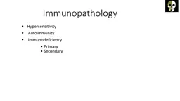
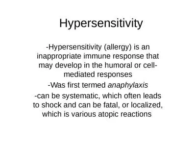
Diagnosis of cellmediated responses 1 Delayed hypersensitivity reactions Scratch skin test Intradermal skin test Patch test 2 Lymphocyte transformation test Lymphocyte activation test ID: 599589

test skin allergic reactions skin test reactions allergic patch minutes allergy nurse the

Share:

Link:

Embed:

Download Presentation from below link

Download Presentation The PPT/PDF document "Diagnosis of cell-mediated responses" is the property of its rightful owner. Permission is granted to download and print the materials on this web site for personal, non-commercial use only, and to display it on your personal computer provided you do not modify the materials and that you retain all copyright notices contained in the materials. By downloading content from our website, you accept the terms of this agreement.


Presentation Transcript

Slide1

Diagnosis of cell-mediated responsesSlide2

Diagnosis of cell-mediated responses

1. Delayed hypersensitivity reactions .

Scratch skin test.

Intradermal skin test

Patch test.

2. Lymphocyte transformation test .

Lymphocyte activation test

. ( detect

markers by flow

cytometry)Slide3

Antibodies, which are circulating 

proteins, respond

 within minutes, to give what, is termed an immediate hypersensitivity reaction. 

T

 cells responses occur over several days, and are thus called delayed hypersensitivity reactions. The cascade of events initiated by the T cells leads to hardening(induration) and redness (erythema) at the injection site.

Delayed Hypersensitivity Skin TestSlide4

Delayed Hypersensitivity Skin Test

Antibodies, which are circulating 

proteins, respond


within minutes, to give what, is termed an immediate hypersensitivity reaction. 

T

 cells responses occur over several days, 

and are

 thus called delayed hypersensitivity reactions. 

The

 cascade of events initiated by the T cells leads to 

hardening(induration

) and redness (erythema) at the injection site.Slide5

Skin testing

Skin testing is usually done at a doctor's office. A nurse generally administers the test, and a doctor interprets the results. Typically, this test takes about 20 to 40 minutes.

Some

tests detect immediate allergic reactions, which develop within minutes of exposure to an allergen

.

Other

tests detect delayed allergic reactions, which develop over a period of several days

.Slide6

Skin prick test

A

skin prick test, also called a puncture or scratch test, checks for immediate allergic reactions to as many as 40 different substances at once. This test is usually done to identify allergies to pollen, mold, pet dander, dust mites and foods. In adults, the test is usually done on the forearm. Children may be tested on the upper back

.

Allergy skin tests aren't painful. This type of testing uses needles (lancets) that barely penetrate the skin's surface. You won't bleed or feel more than mild, momentary discomfort

.Slide7

After cleaning the test site with alcohol, the nurse draws small marks on your skin and applies a drop of allergen extract next to each mark. He or she then uses a lancet to prick the extracts into the skin's surface. A new lancet is used for each allergen.Slide8

About 15 minutes after the skin pricks, the nurse observes your skin for signs of allergic reactions. If you are allergic to one of the substances tested, you'll develop a raised, red, itchy bump (wheal) that may look like a mosquito bite. A nurse will then measure the bump's size

.

After the nurse records the results, he or she will clean your skin with alcohol to remove the marks.Slide9

Quality control

To see if your skin is reacting normally, two additional substances are scratched into your skin's surface:

Histamine.

 In most people, this substance causes a skin response. If you don't react to histamine, your allergy skin test may not reveal an allergy even if you have one.

Glycerin or saline. In most people, these substances don't cause any reaction. If you do react to glycerin or saline, you may have sensitive skin. Test results will need to be interpreted cautiously to avoid a false allergy diagnosis.Slide10

Skin injection

test

You

may need a test that uses a needle to inject a small amount of allergen extract just into the skin on your arm (intradermal test).

The

injection site is examined after about 15 minutes for signs of an allergic reaction.

Your

doctor may recommend this test to check for an allergy to insect venom or penicillin.Slide11

Patch

test

Patch

testing is generally done to see whether a particular substance is causing allergic skin irritation (contact dermatitis).

Patch

tests can detect delayed allergic reactions, which can take several days to develop

.

Patch tests don't use needles. Instead, allergens are applied to patches, which are then placed on your skin.

During

a patch test, your skin may be exposed to 20 to 30 extracts of substances that can cause contact dermatitis. These can include latex, medications, fragrances, preservatives, hair dyes, metals and resins.Slide12

You wear the patches on your arm or back for 48 hours. During this time, you should avoid bathing and activities that cause heavy sweating.

The patches are removed when you return to your doctor's office. Irritated skin at the patch site may indicate an allergy.Slide13

Lymphocyte activation test.

The test is based on the principle of antigen/allergen-specific induction of cell division in lymphocytes following contact with their «fitting« antigen.

A

positive reaction in the LTT indicates the presence of antigen-specific lymphocytes (memory cells) in the patient’s blood

.

In the

p

ast the

LTT was at best equal to the skin test, if not

inferior but today

, the LTT technologies used in

specialized

immunological laboratories are very reliable and offer high sensitivity and specificity

.

In LTT cells markers are determined by flow cytometry.