Megan Mair Langerhans Cell Histiocytosis An Overview Sawyer diagnosed with histiocytosis of the mastoid and skull at age 2 Langerhans Cell Histiocytosis LCH a rare blood disorder characterized by the abnormal development and proliferation of Langerhans Cells within the bo ID: 642642
Download Presentation The PPT/PDF document "Differentiation of LCH-Like Cells" is the property of its rightful owner. Permission is granted to download and print the materials on this web site for personal, non-commercial use only, and to display it on your personal computer provided you do not modify the materials and that you retain all copyright notices contained in the materials. By downloading content from our website, you accept the terms of this agreement.
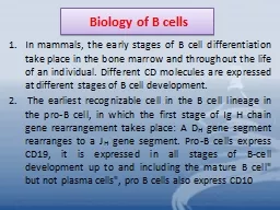
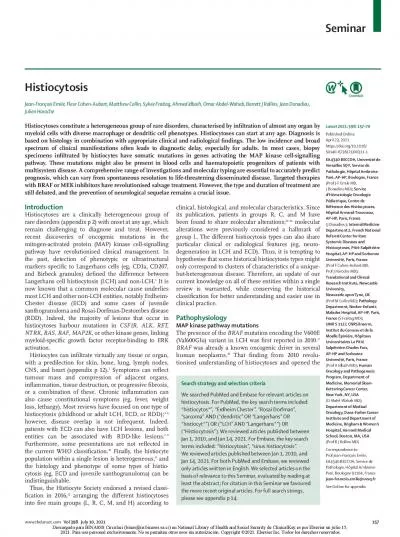
Slide1
Differentiation of LCH-Like Cells
Megan MairSlide2
Langerhans Cell
Histiocytosis: An Overview
- Sawyer: diagnosed with
histiocytosis
of the mastoid and skull at age 2 - Langerhans Cell Histiocytosis (LCH): a rare blood disorder characterized by the abnormal development and proliferation of Langerhans Cells within the body, resulting in cellularly diverse tumorscolloquially known as a “relative of cancer”- Langerhans Cells: self-dividing dendritic cells primarily found on the skin. Origin of the abnormal LCs found in the abnormal locations typical of LCH is unknown. Slide3
Disease
LCH Lesions
Cell
Behavior
LCH CellsCell O
rigin
Disease origin? Breakdown
the partsSlide4
LC-like behavior
LC-like cells
Monocyte CD14+ from blood
Geissmann
et al
.
: proposed origin of LC-like cells
in vitro
LCH-like behavior
LCH-like cells
Monocyte CD14+ from LCH lesion
Hypothesis: same conditions result in development of LCH-like cells
in vitro
Measures of LCH-like molecular behavior:“Cytokine storm”: levels of pro-inflammatory cytokines
interleukin-1α (IL-1α) and interferon-γ (IFN-γ), as well as anti-inflammatory
interleukin-10 (IL-10) and growth factor GM-CSF Lesion formation: irregularities in cell-regulating NF-
κB
pathway, measured through p
resence
of
P16
and
P21
Grown in media with TGFβ1, GM-CSF, and IL-4
Cell origin from monocytes?
Collect samples from blood and lesion
s
ite of LCH patient and sample from
blood of healthy non-LCH patient; expose to same growth conditionsSlide5
1) Levels of IL-1α, IFN-γ, IL-10, GM-CSF
Polymerase
c
hain reaction to amplify level of producing RNAs
4)
Heating/cooling cycles break apart strands and continue to synthesize cDNA between heat cycles, amplifying initial RNA products
1)
Purification from other cell products via
CsCl
gradient. RNA stabilized through
polyadenylation
2)
Total RNA reverse transcribed using enzyme
Moloney
murine leukemia reverse transcriptase (BRL)
3
)
Target cDNA found in PCR cycles via primer sequences. Primer
sequences for cytokines IL-1α, IFN-γ, IL-10,
as well as GM-CSF are available on
GenBank
Procedures described by
Enk
& KatzSlide6
1
)
Amplified RNA products bind to complementary probes labeled by radioactive isotope Phosphorous-32
1) Levels of IL-1α, IFN-γ, IL-10, GM-CSF
2)
Liquid hybridization techniques
2)
Each sample loaded onto PAGE gel and electrophoresed
3
)
Samples dried and analyzed for
bp
signals, and radiation levels analyzed via
densitometric
scanner
Radiation levels versus hours after
hapten
application. Computer-generated graphs measuring radiation levels within each sample. For our proposes, sample type replaces time.Slide7
2) Irregular presence of P16 and P21
Mutation in BRAF V600E gene found in many cases of both melanoma and LCH (
Badalian
-Very)
NF-
κB
: protein complex responsible regulating DNA transcription within animal cells
Irregularities in signaling pathway result in abnormal cell behavior that can lead to tumor formationSlide8
2) Irregular presence of P16 and P21
ELISA kits available for target antigensLabeling antibody emits dye, indicating presence of target antigenSlide9
Potential Outcomes
LCH-like cells can be differentiated from CD14+ monocytes
G
enomic
differences in monocytes found in lesion site are likely accountable for this altered differentiation pattern from regular LCsFurther suggests monocyte CD14+ is involved in initial production of LCs
DNA sequencing of blood and lesion site monocytes can identify differences and potential oncogenes; a similar study by
Fearon
& Vogelstein
found genetic differences in cancer and noncancerous colon cells
Regular LCs are the body’s first defense
against diseases such as HIV; this knowledge may help in developing preventatives and treatments to other autoimmune disorders
LCH-like cells cannot be differentiated from CD14+ monocytes
Further
experimentation
of other
proposed LC differentiation origins via different monocytes can be tested using the same procedures; if no results, it can be assumed there are no differences within the monocyte precursors in LCH cells, indicating non-myeloid environmental causes for the development of the
disease Slide10
References
Badalian-very G,
Vergilio
JA,
Degar BA, Rodriguez-galindo C, Rollins BJ. Recent advances in the understanding of Langerhans cell histiocytosis. Br J Haematol. 2012;156(2):163-72.Badalian-Very, Gayane et al. “Recurrent BRAF Mutations in Langerhans Cell Histiocytosis
.”
Blood
116.11
Enk
, A.H., Katz, S.I.(1992).Early molecular events in the induction phase of contact sensitivity. Proc. Natl. Acad. Sci. USA,89, 1398-1402.
Fearon ER, Vogelstein B. A genetic model for colorectal tumorigenesis
. Cell. 1990;61(5):759-67. Hayworth, Douglas. Overview of ELISA. 2014. Web. http://www.piercenet.com/method/overview-elisa Hoesel
B, Schmid JA. The complexity of NF-κB signaling in inflammation and cancer. Mol Cancer. 2013;12:86. Madonna
G, Ullman CD, Gentilcore G, Palmieri G, Ascierto PA. NF-κB as potential target in the treatment of melanoma. J
Transl Med. 2012;10:53. Valladeau, Jenny; Dezutter-Dambuyant, Colette;
Saeland, Sem (2003). "Langerin/CD207 Sheds Light on Formation of Birbeck
Granules and Their Possible Function in Langerhans Cells". Immunologic Research 28 (2): 93–107.