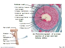
/
 (b) Photomicrograph of a cross  (b) Photomicrograph of a cross

(b) Photomicrograph of a cross - PowerPoint Presentation

alida-meadow
alida-meadow . @alida-meadow
Follow
345 views
Uploaded On 2020-04-04

(b) Photomicrograph of a cross - PPT Presentation

section of a hair and hair follicle 250x Connective tissue root sheath Follicle wall Cuticle Glassy membrane Cortex Medulla Internal epithelial root sheath ID: 775329

hair root skin degree hair root skin degree sheath follicle figure burns nail epithelial burn bulb body tissue gland

Share:

Link:

Embed:

Download Presentation from below link

Download Presentation The PPT/PDF document " (b) Photomicrograph of a cross" is the property of its rightful owner. Permission is granted to download and print the materials on this web site for personal, non-commercial use only, and to display it on your personal computer provided you do not modify the materials and that you retain all copyright notices contained in the materials. By downloading content from our website, you accept the terms of this agreement.


Presentation Transcript

Slide1

(b) Photomicrograph of a cross

section of a hair and hair

follicle (250x)

• Connective tissue

root sheath

Follicle wall

• Cuticle

• Glassy membrane

• Cortex

• Medulla

• Internal epithelial

root sheath

• External epithelial

root sheath

Hair

Hair shaft

Arrector

pili

Sebaceous

gland

Hair root

Hair bulb

Figure 5.6b

Slide2

Hair Follicle

Extends from the epidermal surface into dermis

Two-layered wall: outer connective tissue root sheath, inner epithelial root sheath

Hair bulb: expanded deep end

Slide3

Hair Follicle

Hair follicle receptor (root hair plexus)

Sensory nerve endings around each hair bulb

Stimulated by bending a hair

Arrector pili

Smooth muscle attached to follicle

Responsible for “goose bumps”

Slide4

Figure 5.6c

Hair shaft

Arrector

pili

Sebaceous

gland

Hair root

Hair bulb

(c)

Diagram of a longitudinal view of the expanded hair

bulb of the follicle, which encloses the matrix

Internal epithelial root sheath

External epithelial root sheath

Connective tissue root sheath

Follicle wall

Hair matrix

Melanocyte

Hair papilla

Subcutaneous adipose tissue

Medulla

Cortex

Cuticle

Glassy membrane

Hair root

Slide5

(d) Photomicrograph of longitudinal view

of the hair bulb in the follicle (160x)

Follicle wall

Hair matrix

Hair papilla

Subcutaneous

adipose tissue

Hair root

• Connective

tissue root sheath

• Glassy membrane

• External epithelial

root sheath

• Internal epithelial

root sheath

• Cuticle

• Cortex

• Medulla

Hair shaft

Arrector

pili

Sebaceous

gland

Hair root

Hair bulb

Figure 5.6d

Slide6

Types of Hair

Vellus—pale, fine body hair of children and adult females

Terminal—coarse, long hair of eyebrows, scalp, axillary, and pubic regions (and face and neck of males)

Slide7

Types of Hair

Hair Growth

Growth phase (weeks to years) followed by regressive stage and resting phase (1–3 months)

Growth phase varies (6–10 years in scalp, 3–4 months in eyebrows)

Slide8

Hair Thinning and Baldness

Alopecia—hair thinning in both sexes after age 40

True (frank) baldness

Genetically determined and sex-influenced condition

Male pattern baldness is caused by follicular response to DHT

Slide9

Structure of a Nail

Scalelike modification of the epidermis on the distal, dorsal surface of fingers and toes

Slide10

Figure 5.7

Lateral

nail fold

Lunule

Nail

matrix

Root of nail

Proximal

nail fold

Hyponychium

Nail bed

Phalanx (bone of fingertip)

Eponychium

(cuticle)

Body

of nail

Free edge

of nail

(a)

(b)

Slide11

Functions of the Integumentary System

Protection—three types of barriers

Chemical

Low pH secretions (acid mantle) and defensins retard bacterial activity

Slide12

Functions of the Integumentary System

Physical/mechanical barriers

Keratin and glycolipids block most water and water- soluble substances

Limited penetration of skin by lipid-soluble substances, plant oleoresins (e.g., poison ivy), organic solvents, salts of heavy metals, some drugs

Biological barriers

Dendritic cells, macrophages, and DNA

Slide13

Functions of the Integumentary System

Body temperature regulation

~500 ml/day of routine insensible perspiration (at normal body temperature)

At elevated temperature, dilation of dermal vessels and increased sweat gland activity (sensible perspirations) cool the body

Cutaneous sensations

Temperature, touch, and pain

Slide14

Functions of the Integumentary System

Metabolic functions

Synthesis of vitamin D precursor and collagenase

Chemical conversion of carcinogens and some hormones

Blood reservoir—up to 5% of body’s blood volume

Excretion—nitrogenous wastes and salt in sweat

Slide15

Skin Cancer

Most skin tumors are benign (do not metastasize)

Risk factors

Overexposure to UV radiation

Frequent irritation of the skin

Some skin lotions contain enzymes in liposomes that can fix damaged DNA

Slide16

Skin Cancer

Three major types:

Basal cell carcinoma

Least malignant, most common

Squamous cell carcinoma

Second most common

Melanoma

Most dangerous

Slide17

Basal Cell Carcinoma

Stratum basale cells proliferate and slowly invade dermis and hypodermis

Cured by surgical excision in 99% of cases

Slide18

Figure 5.8a

Slide19

Squamous Cell Carcinoma

Involves keratinocytes of stratum spinosum

Most common on scalp, ears, lower lip, and hands

Good prognosis if treated by radiation therapy or removed surgically

Slide20

Figure 5.8b

Slide21

Melanoma

Involves melanocytes

Highly metastatic and resistant to chemotherapy

Treated by wide surgical excision accompanied by immunotherapy

Slide22

Melanoma

Characteristics (ABCD rule)

A: Asymmetry; the two sides of the pigmented area do not match

B: Border exhibits indentations

C: Color is black, brown, tan, and sometimes red or blue

D: Diameter is larger than 6 mm (size of a pencil eraser)

Slide23

Figure 5.8c

Slide24

Burns

Heat, electricity, radiation, certain chemicals

Burn

(tissue damage, denatured protein, cell death)

Immediate threat:

Dehydration and electrolyte imbalance, leading to renal shutdown and circulatory shock

Slide25

Rule of Nines

Used to estimate the volume of fluid loss from burns

Slide26

Figure 5.9

Anterior and posterior

head and neck, 9%

4

1

/

2

%

4

1

/

2

%

Anterior and posterior

upper limbs, 18%

Anterior and posterior

lower limbs, 36%

100%

Totals

Anterior and posteriortrunk, 36%

Anteriortrunk,18%

9%

9%

(Perineum, 1%)

4

1

/

2

%

Slide27

Partial-Thickness Burns

First degree

Epidermal damage only

Localized redness, edema (swelling), and pain

Second degree

Epidermal and upper dermal damage

Blisters appear

Slide28

Figure 5.10a

(a)

Skin bearing partial

thickness burn (1st and

2nd degree burns)

1st degree

burn

2nd degree

burn

Slide29

Full-Thickness Burns

Third degree

Entire thickness of skin damaged

Gray-white, cherry red, or black

No initial edema or pain (nerve endings destroyed)

Skin grafting usually necessary

Slide30

Figure 5.10b

(b)

Skin bearing full

thickness burn

(3rd degree burn)

3rd

degree

burn

Slide31

Severity of Burns

Critical if:

>25% of the body has second-degree burns

>10% of the body has third-degree burns

Face, hands, or feet bear third-degree burns

Slide32

Developmental Aspects: Fetal

Ectoderm

epidermis

Mesoderm

dermis and hypodermis

Lanugo coat: covering of delicate hairs in 5th and 6th month

Vernix caseosa: sebaceous gland secretion; protects skin of fetus

Slide33

Developmental Aspects: Adolescent to Adult

Sebaceous gland activity increases

Effects of cumulative environmental assaults show after age 30

Scaling and dermatitis become more common

Slide34

Developmental Aspects: Old Age

Epidermal replacement slows, skin becomes thin, dry, and itchy

Subcutaneous fat and elasticity decrease, leading to cold intolerance and wrinkles

Increased risk of cancer due to decreased numbers of melanocytes and dendritic cells