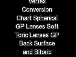
/
Contact Lens Clinical Pearls Pocket Guide  CONTENTS Vertex Conversion Chart Spherical Contact Lens Clinical Pearls Pocket Guide  CONTENTS Vertex Conversion Chart Spherical

Contact Lens Clinical Pearls Pocket Guide CONTENTS Vertex Conversion Chart Spherical - PDF document

alida-meadow
alida-meadow . @alida-meadow
Follow
827 views
Uploaded On 2014-11-23

Contact Lens Clinical Pearls Pocket Guide CONTENTS Vertex Conversion Chart Spherical - PPT Presentation

gpliinfo brPage 3br Vertex Conversion Chart minus plus 387 400 425 400 425 450 425 450 475 450 475 500 475 500 525 500 525 562 512 550 587 537 575 612 562 600 650 575 625 675 600 650 700 625 675 737 650 700 762 662 725 800 687 750 825 712 775 850 725 ID: 15579

gpliinfo brPage 3br Vertex Conversion

Share:

Link:

Embed:

Download Presentation from below link

Download Pdf The PPT/PDF document "Contact Lens Clinical Pearls Pocket Guid..." is the property of its rightful owner. Permission is granted to download and print the materials on this web site for personal, non-commercial use only, and to display it on your personal computer provided you do not modify the materials and that you retain all copyright notices contained in the materials. By downloading content from our website, you accept the terms of this agreement.


Presentation Transcript

1 Contact Lens Clinical PearlsPocket Guide 2 CONTENTS Vertex Conversion Chart Spherical GP Lenses Soft Toric LensesGP Back Surface and Bitoric LensesMultifocal LensesOrthokeratologyKeratoconus Scleral Lens Fitting and HandlingDiopter to Radius (mm) Conversion Chart Special Thanks To the GP Lens Institute Advisory Committeeand especially Stephen Byrnes, Greg DeNaeyer, Tim Edrington,Chris Gilmartin, Bob Grohe, Lynette Johns, John Laurent, Norman Leach, Derek Louie, Miles Press, Jack Schaeffer, Christine Sindt, Eef van der Worp, Michael Ward, and Frank Weinstockwww.gpli.info 3 Vertex Conversion Chart minusplus - 3.87 4.00 +4.25 - 4.00 4.25 +4.50 - 4.25 4.50 +4.75 - 4.50 4.75 +5.00 - 4.75 5.00 +5.25 - 5.00 5.25 +5.62 - 5.12 5.50 +5.87 - 5.37 5.75 +6.12 - 5.62 6.00 +6.50 - 5.75 6.25 +6.75 - 6.00 6.50 +7.00 - 6.25 6.75 +7.37 - 6.50 7.00 +7.62 - 6.62 7.25 +8.00 - 6.87 7.50 +8.25 - 7.12 7.75 +8.50 - 7.25 8.00 +8.87 - 7.50 8.25 +9.12 - 7.75 8.50 +9.50 - 7.87 8.75 +9.75 - 8.12 9.00 +10.12 - 8.37 9.25 +10.37 - 8.50 9.50 +10.75 - 8.75 9.75 +11.00 - 8.87 10.00 +11.37 - 9.37 10.50 +12.00 - 9.75 11.00 +12.75 - 10.12 11.50 +13.37 - 10.50 12.00 +14.00 - 10.87 12.50 +14.75 - 11.25 13.00 +15.50 - 11.62 13.50 +16.12 - 12.00 14.00 +16.75 - 12.37 14.50 +17.50 - 12.75 15.00 +18.25 - 13.00 15.50 +19.00 - 13.50 16.00 +19.75 - 13.75 16.50 +20.50 - 14.12 17.00 +21.50 - 14.50 17.50 +22.25 - 14.75 18.00 +23.00 - 15.12 18.50 +23.75 - 15.50 19.00 +24.75 4 SPHERICAL GP LENSESBefore the FitPresent adaptation with terms such as “lens awareness” and “lid sensation.”Use a topical anesthetic prior to initial lens application.Empirical versus Diagnostic FittingDiagnostic fittinghas the benefit of obtaining an optimumfit and power through the application of lenses.Empirical fittinghas the benefit of often having the first experience with GP lenses be a very positive one visually.Fitting PearlsUse a cobalt blue filter in combination with a yellow (Wratten) filter for optimum evaluation of the fluorescein pattern.Strive for an alignment fit. This is often achieved with a base curve radius (BCR) equal to the flatter K reading (termed “on K”) or slightly flatter than K due to the asphericity of the cornea.Increasing the optical zone diameter(OZD) increases sagittal depth and effectively steepens the fit. Decreasing the OZDdecreases sagittal depth and effectively flattens the fit.Flattening the peripheral curve radius (PCR) and/or increasing the curve width will increase edge clearance. Steepening the PCR and/or reducing the curve width will decrease edge clearance.When you make a design change, make it a significant one. Alignment Fit 5 Change the base curve by ≥0.50D; OAD/OZD ≥ 0.3mm; center thickness ≥0.03mm; peripheral curve radius/width ≥1.0/0.2mm.Order a plus lenticular to reduce edge thickness on all high minus 5.00D) and a minus lenticular on all plus and low minus (1.50D) lenses to increase edge thickness.The use of a lenticular, when indicated, in combination with an ultrathin center thickness, will reduce the risk of inferior decentration.To predict the lens power, take the spherical refractive value at the corneal plane. If selecting a BCR flatter than K, add the corresponding amount of plus power. If fitting steeper, add minus power (SAM/FAP or steep add minus, flat add plus). Ignore the cylinder power and the steeper K value. Example: Rx 4.50 0.75 x 180; K’s 43.00 @ 180/ 43.75 @ 090 BCR 42.50D (0.50D flatter than KThe predicted power is 4.25D (4.50D at the corneal plane) + 0.50D (FAP) = 3.75DResourcesResources on fitting and problemsolving GP lenses are available at www.gpli.info. OFT TORIC LENSESBefore the FitPatients with as low as 0.75D refractive cylinder are often good candidates.If a patient has ≥ 0.75D cylinder and is wearing a spherical lens, provide them with a toric lens to compare the quality of vision.One method of determining how good a candidate someone is for soft toric lenses is the “Becherer Twist.” With the patient’s subjective refraction in the phoropter, twist the cylinder axis knob until the acuity chart first blurs. If the twisting is 20˚ in each direction, 90% of the patients will be successful. If it blurs at 15˚, 90% success is achieved with two lenses. At 10˚, 70% will be Spherical GP Lenses cont’d 6 successful with 3 lenses. If there is blur with 5rotation, success is unlikely.The Fitting ProcessSelect diagnostic lenses as close to the refractive power at thecorneal plane as possible.Example: a patient witha refraction of: 4.75 1.50 x 180 should be fit with adiagnostic lens of 4.50 1.25 x 180 (the powers at thecorneal plane). If the exact cylinder power is not available, select thelower cylinder power lens. For instance, if the patient has 1.50 x 180cylinder power at the corneal plane and the soft toric lens cylinder isavailable in either 1.25 or 1.75 x 180, select the 1.25 x 180lens.Allow the lens to settle for a minimum of 10 minutes prior to evaluation.To determine the amount of lens rotation, use a degree scale on the biomicroscope to line up an optic section with the axis mark on the lens. With this method, an exact amount ofrotation can be read directly off the degree scale. If this scale is not available, the clockapproach can be used to estimate theamount of rotation. With this method, each hour on a clock is equivalent to 30To determine the axis of the final lens, use the “left add, right subtract” (LARS) technique. If the patient has a refraction of 2.00 1.25 x 180, and a lens with these identical parameters is applied to the right eye and rotates 10˚ to the observer/practitioner’s right (nasally), a lens with an axis of 170˚ should be fit. If it rotates 10˚ to the observer’s left (temporally), a lens with a 10˚ axis should be fit.Subtract the amount of rotation from the prescription, not the diagnostic lens. Also, the final lens ordered should fit in the same skewed position as the diagnostic lens. Rx axis at 180° and no rotation; OrderLens Axis at 180° Rx axis at 180° and 10° right rotation; Order Lens at Axis 170° Rx axis at 180° and 10° left rotation; Order Lens at Axis 010° oft ToricLenses cont’d 7 Poor Visual Responsef reduced vision is present with a spherical overrefraction (OR), a spherocylindrical OR should be performed. To determinethe recommended cylinder power and axis based upon the OR, useone of many available cross cylinder calculators. These may be found on the AOA Contact Lens and Cornea Section web site, www.eyedock.com, as well as many softlens company web sites. If the patient experiences a persistent problem with vision due to lens rotation or other reasons, a GP lens should be considered.GP BACK SURFACE AND BITORIC LENSES Before the FitGood candidates are individuals wit�h 2.00D of corneal cylinder who do not achieve good centration and/or have a poorly aligned fit, are dissatisfied with their vision from soft toric lenses, or have critical vision needs.DecisionMaking Process A back surface toric lens induces a cylinder equal to almost half of the back surface toricity with the same axis (the exact amount depends upon the refractive index of material). When the cylinder is corrected on the front surface, it results in a bitoric lens. A back toric design (spherical front surface) isrecommended when the residual astigmatism is approximately one half of the back surface toricity and has an axis equal to the flat K reading. This should also be considered when the patient’s refractive cylinder is approximately 1½ times the corneal cylinder. Empirical Versus Diagnostic FittingDiagnostic fittinghas the benefit of obtaining an optimum fit and power through the application of lenses. Diagnostic fitting sets are oft ToricLenses cont’d 8 typically available from your laboratory. 3.00D toricity sets are especially popular.Empirical fittinghas the benefit of often having the first experience with GP lenses be a very positive one visually. MandellMoore and the Quinn GP Toric calculators are simple and successful methods of calculating the base curve radii and powers based on the refraction and K values. These guides are available www.gpli.infoFitting PearlsBitoric lens power determination only differs from spherical lenses in that two tear lens power calculations are needed, not one.The flatter base curve is typically selected approximately 0.25D flatter than flat K. The steeper base curve radius is typically 1.00D flatter than steep K. This creates 0.75D of toricity, which would simulate the ideal cornea to fit a spherical lens. The result should be an alignment fitting relationship. If a diagnostic lens is selected, a spherical overrefraction should be performed and the resulting power should be added to the power in each meridian. If the diagnostic lens powers are plano in tmeridian and 3.00D in the 90˚ meridian and the overrefraction is 1.00D, the final powers are 1.00D and 4.00D. This lens can rotate on the eye without affecting vision (spherical power effect).If the patient has residual astigmatism, it is likely that a spherocylindrical overrefraction will result in optimum acuity. If so, the overrefraction in one meridian should be added to the diagnostic lens power in that meridian. In the above example, if the overrefraction is 1.00 1.00 x 180, 1.00D would be added to plano, and 2.00D (in the 90˚ meridian) would be added to 3.00D resulting in powers of .00D / 5.00D.Toric GP lenses are not often indicated with irregular cornea patients as the refractive axis differs from the corneal axis and bitoric designs have base curve radii and corresponding powers 90˚ apart. GP Back Surface & Bitoric cont’d 9 MULTIFOCAL LENSESDecisionMaking ProcessExplain all options to every presbyope or emerging presbyope including single vision lenses/reading glasses, monovision, and multifocallenses.The benefits of multifocals versus monovision include the quality of vision at all distances out of both eyes. Many comparison studies between soft and/or GP multifocals versus monovision have resulted in approximately 75% of the patients preferring multifocals.Monovision is indicated when the patient either has miotic pupils therefore very little power effect is obtained from the paracentral aspheric region or pupils larger than 6mm, which will result in visual disturbance from the miperiphery of the lens, and does not have critical vision needs.Determine what the patient’s goals are from these lenses. Have the patient rank the importance of distance, intermediate, and near vision and by concentrating on the two most important, the patient will likely be satisfied with the vision at all distances with multifocal lenses. But, it is best to not make any guarantees! Patients need to be given realistic expectations. Most contact lens multifocal designs utilize the simultaneous vision principle in which multiple corrections are in front of the eye at the same time. Some blur may be noted when compared to spectacles. In addition, it may take a lens exchange or two to achieve a successful fit and acceptable vision. Finally, patients should never be told that they will not require spectacles. Some multifocal wearers appreciate a low minus overcorrection when driving while others may desire some additional plus for reading fine print.If they are patient and motivated they will most likely be successful. 10 The Fitting ProcessImportant tests to perform include pupil size measurement, tear film evaluation, and determining lower lid position. When possible, it is important that the patient’s first experience in multifocal lenses is with lenses in their prescription so use lenses from a soft lens inventory or empirically fit GP multifocals.If, after performing visual acuities at both distance and near, vision is reduced out of one eye at aparticular distance, overrefract monocularly with hand held trial lenses or a flipper bar and with both eyes open.When assessing vision at near, make sure the patient is reading material (magazine, book, etc.) that is similar in font to their everydaymaterial and consistent with what they desire to see well at near.Once the lenses have been evaluated, have the patient walk around the office and perform normal visual tasks. Encourage binocular viewing. Discourage viewing monocularly (the “contact lens salute”).If satisfactory vision and fitting relationship is achieved, schedule an appointment, at minimum, one week later to allow the patient to become adapted to the lenses.Soft Multifocal LensesGood candidates include individuals who do not have critical vision demands, have ≤ 0.75D refractive cylinder (unless using toric multifocals), and are satisfied spherical soft lens wearers. If they decide to be fit with soft multifocals, they should be informed about the GP multifocal option if vision becomes problematic.For the initial diagnostic lens, select the power to be equal to the spherical equivalent of the manifest refraction at the corneal plane (unless otherwise recommended by the manufacturer). For example, if the refraction at the spectacle plane is 4.50 0.50 x 180, the initial diagnostic lens would be equal to 4.50D. Multifocal Lenses cont’d 11 The final lens power should be the lens that emphasizes “least minus, most plus” if there is a range of lenses that provide acceptable distance vision.Allow the lenses to settle at least 10 minutes after application before evaluation.Be sure to consult the manufacturers’ fitting guides and problem solving recommendations for their unique lens design. GP Multifocal LensesThis is a viable option for patients with critical vision demands. Aspheric lensesAspheric lenses are a viable option except foindividuals who have a large (≥ 6mm) pupil diameter. For uninterrupted vision at distance and near, segmented translating (bi) multifocal lenses are recommended. Aspheric lenses should result in an alignment fitting relationship, good centration, anlimited (1mm) movement with the blink. If the lens moves excessively, select a lens with a 0.50D steeper base curve radius.Segmented lensesSegmented, translating bifocal lenses typically have the seg line positioned at or slightly below the lower pupil margin. Many designs have an aspheric or segmented intermediate zone, which should be positioned right above the lower pupil margin. The lens should move minimally (1mm) with the blink. Poor candidates for this design would include anyone with a lowpositioning lower lid.When viewing through the biomicroscope, have the patient view inferiorly and the lens should shift upward or translate as the edge contacts the lower lid. If the lens does not translate (or only does so intermittently), increase edge clearance with a flatter base curve radius or peripheral curve radius. Multifocal Lenses cont’d 12 If the lens moves excessively, increase the prism ballast. For assistance with problemsolving GP bifocal and multifocal lens designs, the Contact Lens Manufacturers Association (CLMA) member laboratory consultants are an invaluable resource. In addition, the GP Lens Institute has numerous resources, like the onepictured here, available atwww.gpli.info ORTHOKERATOLOGYBefore the FitGood candidates include 5.00D myopia, ≤ 1.50D WTR corneal cylinder or ≤ 0.50D ATR cylinder; < 6mm pupil diameter. Important screening tests include refraction, slit lamp valuation, and corneal topography. Topography will rule out patients with irregular corneas as well as provide corneal eccentricity values. The Fitting ProcessThe “Jessen formula” can often be used to determine the base curve radius. It uses the FAP (flat add plus) tear lens factor to result in a final power of +0.75D, which will allow for regression during the day. If the patient has a refractive error of 3.00 0.75 x 180 and keratometry values equal to 44.00 @ 180/ 44.75 @ 090, tbase curve should equal 3.75D (3.00D + 0.75D) flatter than K, which is equal to 44.00D 3.75D or 40.25D. The initial diagnostic lens is selected in an effort to achieve a bull’s eye fluorescein pattern (central bearing, paracentral pooling, midperipheral bearing, and slight peripheral clearance). Wait at least 10 to 15 minutes before evaluating the fit. Good centration with minimal (≤ 1mm) lag with the blink is desired.The patient should be evaluated the morning after dispensing.Assess the lens to cornea fitting relationship. Multifocal Lenses cont’d 13 The lenses should be removed to assess corneal integrity. If coalesced corneal staining is present, the lens is too flat centrally. Improvement in unaided visual acuity should be present.Corneal topography should be performed and a bull’s eye pattern (central flattening, paracentral steepening) should be present. If superior flattening is present with an inferior arc of steepening (“smiley face”), the lens is too flat. If little change is present with slight central steepening (“central island”), the lens is too steep. If no obvious topography pattern is present, the patient should wear the lenses for 2 more days and be evaluated. On average, it takes 10 days to reach the treatment goal although it will likely be less for lower myopic and higher for more moderate myopic powers.Provide the patient daily disposable lenses of gradually decreasing power to wear during the treatment period and evaluate at one week. At the end of the treatment period, the lenses should be worn on a retainer basis. This can be anywhere from every night for higher myopic patients to once a week for very low myopes.Patients can selfmonitor retainerwear. Whenever the distance vision becomes blurred, they can wear the lenses overnight. Applying a highly viscous artificial tear prior to inserting the lens has been found to optimize centration and lessen corneal staining. Lens removal should not occur immediately upon awakening. Rewetting drops should be applied before removal and the lower lid margin can be used to gently nudge the lower lens edge to break suction if present. Further expertise through certification and examinations are available at www.paragoncrt.com and www.bausch.com as well as from the Orthokeratology Academy of America at www.okglobal.org. Orthokeratologycont’d 14 KERATOCONUSBefore the FitHallmark clinical signs of keratoconus are Fleischer’s ring and Vogt’s striae.In keratoconus, the steepest area of the corneal topography istypically� 48.00D. Also, if the eccentricity value is greater than or equal to 0.8, it is likely keratoconus.In a moderate to advanced keratoconic patient, in the absence of acorneal topographer, the use of a +1.25D trial lens over thepatient’s side of the keratometer will extend the range by about 8.00D. A +2.25D trial lens will extend the range by approximately 14.00D. The Fitting ProcessIf the apex of the cone is relatively small and centrally located, a traditional small diameter keratoconic lens can be used. If a large oval or globus cone is present or the apex is decentered inferiorly, intralimbal, scleral, piggyback, or hybrid designs have all been successful. With most designs, minimal apical clearance or mild touch is desired. This “three point touch” or bull’s eye fluorescein pattern is most likely achieved on a relatively wellcentered apex.Gross apical bearing can result in corneal staining and possibly scarring. Excessive apical clearance can result in peripheral seal off.The presence of excessive inferior edge clearance can be remediated with designs that allow the inferior edge to tuck in (flatsteep, ACT, quadrant specific). Excessive Apical Clearance Excessive Apical Bearing Three Point Touch 15 A piggyback design combination should be considered if a GP lens alone results in either poor centration, less than optimum comfort, or if scarring is present. A very low power (0.50D) silicone hydrogel soft lens can be placed under the GP. Due to the lens combination it is important that a hyper Dk (�100) GP material be used. If the patient has a low corneal apex resulting in the GP lens positioning low he soft lens, the use of a moderate plus power (+6.00D) soft lens may help the GP lens center due to the thicker center of the soft lens. This should result in little to no change in the GP power as a soft lens contributes only about 20% of its power when used in a piggyback system. For assistance with keratoconic patients, communicate with your CLMA member laboratory consultant. For resources contact the National Keratoconus Foundation www.nkcf.org) and the GP Lens Institute (www.gpli.info).SCLERAL LENS FITTING AND HANDLINGDefinition and ApplicationsScleral lenses can be divided into the following categories based upon overall diameter: Corneoscleral (12.9mm to 13.5mm)Semiscleral (13.6mm to 14.9mm)Miniscleral (15.0mm to 18.0mm)Scleral (�18.0mm) Scleral lenses are indicatedin cases of irregular corneas although corneoscleral lenses have been recommended for healthy cornea patients and scleral lenses have been successful for patients with dry eye syndrome and scarred, severely pathologicacorneas. Lens Handling and Patient EducationLens Insertion (nonfenestrated): Keratoconuscont’d 16 For initial fitting evaluation, the lens should be completely filled with isotonic, preserved artificial tears (e.g. OPTIVE™ Sensitive, Unisol 4). Fluorescein from a strip should be added to the filled bowl. The lens can be supported on a large DMV scleral suction cup or equivalent. Alternatively, a tripod made up of the thumb, middle, and index finger, can be used.The face should be parallel to the ground and the lids must be retracted and wellcontrolled. The patient should look straight down toward the ground. The patient should retract the upper lid while the practitioner retracts the lower lid and raise the lens onto the eye in one continuous motion. The lids should be released prior to lowering the supporting suction cup. If seated, cover the patient’s lap withpaper towels before insertion as some of the solution and fluorescein will overflow and could stain clothing. If a large bubble is observed after insertion, either the lens was not inserted in one continuous motion or the lens well was not completely filled with solution. Remove the lens and reinsert. Lens Removal: As the lens will likely be suctioned, always loosen the lens prior to removal. An appropriate rewetting drop should be applied and the inferior periphery of the lens should be gently pushed in a repeated motion for several seconds.With the superior lid well controlled, the inferior lid can be used to lift the lower portion of the lens away from the eye.Alternatively, a medium DMV suction cup can be used. If so, it should be applied to the inferior lens periphery and then pulled in a direction that is down and out with the removal force directed perpendicular to the lens surface, not along the visual axis.Fitting PrinciplesIt is important for the lens to completely vault the cornea while aligning the lens to the bulbar conjunctiva.Choose the Overall Diameter In general, larger lenses can hold more fluid in the corneal chamber and tend to be more forgiving for the fitter, allowing for more clearance over the cornea. Scleral Lens Fitting & Handlingcont’d 17 Smaller lenses must more closely vault the cornea and demand a more precise central fit. For the most irregular corneas, choose a larger lens diameter (miniversus semior corneoscleral diameter when possible). In addition, some manufacturers provide guidelines for selecting an overall diameter based on horizontal visible iris diameter. Choose an Initial Trial LensFollow manufacturer’s fitting guide or try a more simple approach.Standing beside the patient, look at the corneal profile. If very steep, choose a steeper base curve. If flat, choose a flatter base curve. If average, choose an average base curve. These lenses are fit on the basis of sagittal height, so this method can be very effective when used properly. Examine the Corneal Fit With white light and an optic section at high illumination and medium magnification, set the slit lamp housing off axis and examine the central corneal clearance.You will see severallayers in cross section. The outermost band (dark black) is the lens. The dark area is straddled by two hairline reflections that arise from the front and back surface of the lens. Compare this black layer to the tear lens (green). For example, if the trial lens is known to have a thickness of 300 microns and the tear lens appears to be half that thickness, then the lens vaults the cornea by approximately 100 to 150 microns, which is ideal clearance although this varies by design and is often less if the lens is fenestrated. Apply trial lenses until an acceptable central clearance value has been achieved. Note that after applying any type of scleral lens, it will settle into the conjunctiva over a 30 to 40 minute period. This will decrease the corneal vault and Scleral Lens Fitting & Handlingcont’d 18 possibly lead to touch in an area that was vaulted upon initial application of the lens. A trial lens that shows gross, excessive vaulting of the central cornea initially should be removed and replaced with a flatter base curve. However, if the corneal vault is only mildly excessive upon initial application of the trial lens, it is best to allow the lens to settle since it may yield an ideal corneal vault after 30 to 40 minutes.Corneal versus Peripheral Fitting RelationshipOverall, the fit of a scleral lens can be divided into two parts; the central fit (over the cornea called the “corneal chamber”) and the peripheral fit (over the conjunctiva). Examine the entire corneal chamber under diffuse cobalt blue and high illumination. Note any areas of bearing just as with a corneal GP contact lens. When fitting an irregular cornea, it is common to observe touch or bearing in the midperipheral or peripheral cornea once acceptable central clearance has been obtained. In these cases, additional clearance must be created in the problem area without grossly increasing the central clearance. A reverse geometry design can be employed to vault over the areas of touch/bearing, but compensatory flattening of the base curve must be done to avoid excessive central clearance. Good Fit Down Gaze Good Fit Optic Section Good Fit Scleral Compression Scleral Lens Fitting & Handlingcont’d 19 The peripheral portion of the lens should align with the bulbar conjunctiva. Impingement occurs when there is compression or focal blanching of blood vessels, which can occur anywhere on the scleral haptic, not just the edge.Compression, or general indentation of the conjunctiva, hether at the edge or midperiphery of the lens, may result in seal off, suction and indentation. When blanching occurs, flatten the peripheral curve associated with the area of blanching. Excessive movement and/or bubble formation after lens insertion may indicate the peripheral curve(s) are too loose; therefore, tighten the peripheral curve(s). OvertopographyIt is beneficial to perform computerized topography over the contact lens in situ after it has settled for a few minutes. This can reveal any lens flexure. More than about 0.50D of toricity can be significant and should be addressed by increasing the center thickness if it interferes with vision. Check for Tear ExchangeBefore a scleral lens is dispensed, proper tear exchange must demonstrated. Apply the lens without fluorescein in the filling media. After the lens has been properly applied, instill a generous amount of fluorescein dye over the top of the lens with a dye strip. Periodically examine the tear lens and check for dye thathas made its way behind the lens into the tear chamber. After several minutes, there should be at least a small amount of dye in the tear lens.Tear exchange does not need to be rapid, but it is critical for a proper fit. If in the test for tear exchange there is no fluorescein seen in the corneal chamber after waiting for several minutes, flatten the peripheral fit or increase the overall diameter. Scleral Lens Fitting & Handlingcont’d 20 Diopter to Radius (mm) Conversion Chart DiopterRadiusDiopterRadius 34.00D 9.92mm 44.00D 7.67mm 34.25D 9.85mm 44.25D 7.63mm 34.50D 9.78mm 44.50D 7.58mm 34.75D 9.71mm 44.75D 7.54mm 35.00D 9.64mm 45.00D 7.50mm 35.25D 9.57mm 45.25D 7.46mm 35.50D 9.50mm 45.50D 7.42mm 35.75D 9.44mm 45.75D 7.38mm 36.00D 9.37mm 46.00D 7.34mm 36.25D 9.31mm 46.25D 7.30mm 36.50D 9.24mm 46.50D 7.26mm 36.75D 9.18mm 46.75D 7.22mm 37.00D 9.12mm 47.00D 7.18mm 37.25D 9.06mm 47.25D 7.14mm 37.50D 9.00mm 47.50D 7.11mm 37.75D 8.94mm 47.75D 7.07mm 38.00D 8.88mm 48.00D 7.03mm 38.25D 8.82mm 48.25D 6.99mm 38.50D 8.76mm 48.50D 6.96mm 38.75D 8.70mm 48.75D 6.92mm 39.00D 8.65mm 49.00D 6.89mm 39.25D 8.60mm 49.25D 6.85mm 39.50D 8.54mm 49.50D 6.82mm 39.75D 8.49mm 49.75D 6.78mm 40.00D 8.44mm 50.00D 6.75mm 40.25D 8.39mm 50.25D 6.72mm 40.50D 8.33mm 50.50D 6.68mm 40.75D 8.28mm 50.75D 6.65mm 41.00D 8.23mm 51.00D 6.62mm 41.25D 8.18mm 51.25D 6.58mm 41.50D 8.13mm 51.50D 6.55mm 41.75D 8.08mm 51.75D 6.52mm 42.00D 8.04mm 52.00D 6.49mm 42.25D 7.99mm 52.25D 6.46mm 42.50D 7.94mm 52.50D 6.43mm 42.75D 7.89mm 52.75D 6.40mm 43.00D 7.85mm 53.00D 6.37mm 43.25D 7.80mm 53.25D 6.34mm 43.50D 7.76mm 53.50D 6.31mm 43.75D 7.71mm 53.75D 6.28mm