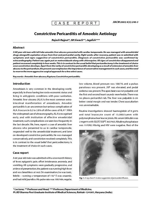
PDF-Fig. 1 : X-ray chest P-A view suggestive of pericardial effusion. ...

Author : alida-meadow | Published Date : 2016-03-08

JIACM 2003 43 2403 Lecturer Professor and Head Professor Department of Medicine Rajesh Rajput SB Siwach JagdishAbstractA 60 year old man with left lobe amoebic

Presentation Embed Code

Download Presentation

Download Presentation The PPT/PDF document "Fig. 1 : X-ray chest P-A view suggestive..." is the property of its rightful owner. Permission is granted to download and print the materials on this website for personal, non-commercial use only, and to display it on your personal computer provided you do not modify the materials and that you retain all copyright notices contained in the materials. By downloading content from our website, you accept the terms of this agreement.

Fig. 1 : X-ray chest P-A view suggestive of pericardial effusion. ...: Transcript


JIACM 2003 43 2403 Lecturer Professor and Head Professor Department of Medicine Rajesh Rajput SB Siwach JagdishAbstractA 60 year old man with left lobe amoebic liver abscess. using LBC Technology. Jong Yull Kim, Prof.,PMIAC. Eulji University. Cellntech Bio.,Co.,Research Institute. Zhanar Yeleubayeva, MD.,MIAC. 1. Kinds of Effusion using LBC Technology. Pleural fluid. Ascitic . tamponade. Sunitha. Daniel. Outline. Brief Overview. Causes. Clinical Presentation. Investigations. Management Update. Overview. Abnormal . amount of and/or an abnormal character to fluid in the pericardial space. SWATI VARA. Contents. How X-rays work . Radiation. Indications . DR. ABCDEFGHI. Common conditions. References. How do X-rays work?. X-rays . are.  electromagnetic . waves. Bone, fat, muscles and . tumours. Fluids . Lab 11, 12. Introduction. Serous fluids are fluids within the closed cavities of the body. These cavities are lined by an adjacent membrane, which forms a double layer of . mesothelial. cells, called the serous membrane. . Presented by :. Dr. . Vidhu. . Mittal. Junior Resident. Deptt. . of Chest and TB. 1. PLEURAL . SPACE. Introduction. The pleural space is a potential space around the lungs, bounded by two membranes, the visceral pleura covering the lung and the parietal pleura covering the chest wall and the diaphragm. . Staff Members of Cardio-thoracic Surgery Departments. Egypt. CARDIAC . TUMOURS. Types:. Primary . Benign (75%): . Myxoma. , . rhabdomyoma. , . fibroma. , . lipoma. , . teratoma. , Papillary . fibroelastoma. DEFINITION. Is an acute inflammation of lung parenchyma caused by various micro organism. Pneumonitis is a general term that describe an inflammatory process in the lung tissue that may predispose or place the at risk for microbial invasion.. Agenda. Effective online feedback: What does the research say? . Feedback in Canvas discussions. Feedback in Canvas quizzes. Feedback in Canvas assignments. Performance task: Create an assignment with corrective, suggestive, and epistemic feedback. . Sheetu. Singh. Assistant Professor. Institute of respiratory disease, . SMS Medical college, . Jaipur. Basics Chest Xray: Dr Sheetu Singh. Chest X Ray. How will you evaluate a chest X ray?. Type of view. Case ReportWe evaluated a 22-year-old patient complaining of acute breathlessness and pretibial edema in relapsed and refractory acute lymphoblastic leukemia (ALL). The patient was under rituximab-cyc Etiology. Idiopathic. Infectious. . • Virus- Coxsackievirus B group . • Bacteria-pneumococci. • Fungus- histoplasma. Noninfectious. • . Uremia. Neoplasm. • Trauma. AUTOIMMUNE. /. HYPERSENSITIVITY. - . fokus. . på. . behandling. . Hjörtur. . Kristjánsson. Klinisk. . presentasjon. . av. . perikardiale. . sykdommer. :. Acute and recurrent pericarditis. Pericardial effusion without major hemodynamic compromise. A 67-year-old man presents to the emergency department . with a 5-day history of fever and cough that produces green sputum. . He . has a history of tobacco use and ischemic cardiomyopathy with . a . Tapping Into Our Shared Experience. Leyla Allum ARNP, Molly Sumner ARNP, Kenneth Rudberg MHA, Aarti Bhat MD. 11/19/2020. Background. Pericardial effusion (PE) and post-pericardiotomy syndrome (PPS) are frequent and well known post-operative complications in patients who have undergone cardiac surgery.

Download Document

Here is the link to download the presentation.
"Fig. 1 : X-ray chest P-A view suggestive of pericardial effusion. ..."The content belongs to its owner. You may download and print it for personal use, without modification, and keep all copyright notices. By downloading, you agree to these terms.

Related Documents