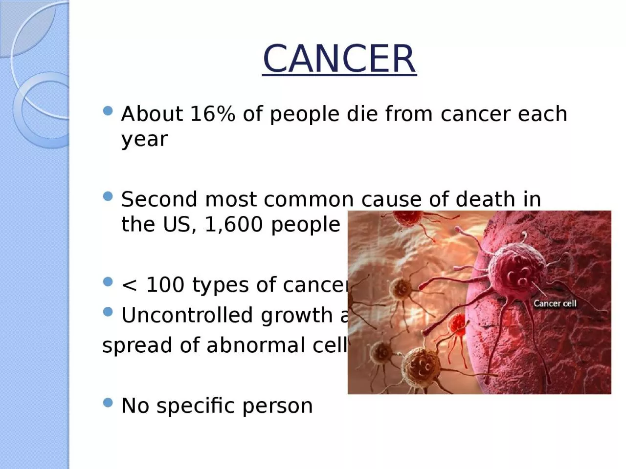
/
CANCER About 16% of people die from cancer each year CANCER About 16% of people die from cancer each year

CANCER About 16% of people die from cancer each year - PowerPoint Presentation

alis
alis . @alis
Follow
66 views
Uploaded On 2024-01-03

CANCER About 16% of people die from cancer each year - PPT Presentation

Second most common cause of death in the US 1600 people per day lt 100 types of cancer Uncontrolled growth and spread of abnormal cells No specific person What is TP53 Better known as p53 ID: 1037337

cancer p53 www senescence p53 cancer senescence www apoptosis pmc cell ncbi nlm nih gov cellular cells articles tumor

Share:

Link:

Embed:

Download Presentation from below link

Download Presentation The PPT/PDF document "CANCER About 16% of people die from canc..." is the property of its rightful owner. Permission is granted to download and print the materials on this web site for personal, non-commercial use only, and to display it on your personal computer provided you do not modify the materials and that you retain all copyright notices contained in the materials. By downloading content from our website, you accept the terms of this agreement.


Presentation Transcript

1. CANCERAbout 16% of people die from cancer each yearSecond most common cause of death in the US, 1,600 people per day< 100 types of cancerUncontrolled growth and spread of abnormal cellsNo specific person

2. What is TP53? Better known as p53Codes for the regulation of the cell cycle preventing the progression of a tumor P53 protects the integrity of the cell by regulating several cellular processes when triggered by stress signals

3. Possible Mediators Several different ways p53 can be activatedDifferent mechanisms by which the tumor can be suppressed

4. Cellular Senescence?Irreversible arresting of cell proliferationFirst studied by Hayflick and Moorhead in the 1960sQualitatively equal to apoptosis in that it prevents the progression of cell proliferation.Cells that are Pro-senescent are actively anti-apoptotic, and senescent cells are resistant to apoptosis.

5. Hirao et al Investigated the activation of p53 using Kinase Chk2.They concluded that after exposing the cells to radiation there was an increase in levels of p53.

6. Taking a step further…Instead of just activating p53 we want to see what mechanism it is more likely to be mediated.

7. Central Question What response is p53 mediating, more frequently, in order to suppress a tumor or DNA damage, APOPTOSIS OR CELLULAR SENESCENCE?

8. Overview of the ExperimentGoal: What tumor suppressor mechanism is happening in most occurrences of a tumor How? Using ionizing radiation & biomarkers

9. How to begin…Obtaining Cancerous cells -Can be made artificially or collected from cancer patients Similar stages in development for comparative purposes, if not the same patientDale et al work with susceptibility

10. Ionizing Radiation Damages the DNA using UV light, removing electrons from the atom, disrupting the DNAIons physically break the base pairs of the DNA

11. Ionizing Radiation Damage is dependent on the dose. The dose must be enough to damage the DNA, but not too strong that it destroys the nucleus. Jiang et al

12. Sa-ß-Gal Senescence-associated beta-galactosidase Glycoside Hydrolase enzyme Found in only senescent cellsWhen in contact with comes in contact with X-gal cleaves the Glycosidic bond and 5-bromo-4-chloro-3-hydroxyindole

13. Cytochemical AssayIncubated with X-gal bufferRinse with Phosphate Buffer SolutionCount occurrences of blue stainsUse of staining to identify biochemical contents in a cell

14. Release of Cytochrome CPrimarily in the mitochondria Protein for life-support function of ATP synthesisA trigger for apoptosis but when found in the cytosol is telling that apoptosis has occurred.

15. CentrifugeTissue Homogenizer Three Rounds, Three different speeds, Three time intervals -Under cold temperature -Removing Supernatant

16. Western BlottingElectrophoresis, with PBS, using the third Cytochrome C antibodiesStain in each well for detection

17. LimitationsIonizing Radiation may lead to apoptosisp53 may not activate p53 may not induce either Cellular Senescence or Apoptosis. The tumor may not be effectively suppressed

18. ConclusionLooking to see that Cellular Senescence is more prevalent.Senescence is a more protective mechanism These cells are still stably viable May have the ability to remodel cells favorably Could lead to effective breakthroughs in cancer research If we are able to manipulate cellular senescence more fitting result for cancer therapies

19. References: 1. “Activating p53 in Cancer Cells with Protein Therapy Shows Preclinical Promise.” PLoS Biology 2.2 (2004): e58. PMC. Web. 26 Nov. 2017. https://www.ncbi.nlm.nih.gov/pmc/articles/PMC340958/ 2. A W Abu-Qare, M B Abou-Donia. “Biomarkers of Apoptosis: Release of Cytochrome c, Activation of Caspase-3, Induction of 8-Hydroxy-2'-Deoxyguanosine, Increased 3-Nitrotyrosine, and Alteration of p53 Gene.” Sigma-Aldrich: Analytical, Biology, Chemistry & Materials Science Products and Services., Journal of Toxicology and Environmental Health. Part B, Critical Reviews, 16 Aug. 2001, www.sigmaaldrich.com/catalog/papers/11503418.3. Brooks, Christopher L, and Wei Gu. “New Insights into p53 Activation.” Nature, 20 Apr. 2010, https://www.nature.com/articles/cr2010534. Childs, Bennett G et al. “Senescence and Apoptosis: Dueling or Complementary Cell Fates?” EMBO Reports 15.11 (2014): 1139–1153. PMC. Web. 26 Nov. 2017. https://www.ncbi.nlm.nih.gov/pmc/articles/PMC4253488/ 5. Demaria, Marco, et al. “Cellular Senescence Promotes Adverse Effects of Chemotherapy and Cancer Relapse.” Cancer Discovery, American Association for Cancer Research, 1 Feb. 2017, cancerdiscovery.aacrjournals.org/content/7/2/165.6. Dimri, Goberdhan. “What Has Senescence Got to Do with Cancer?” Cancer Cell, Cell Press, 13 June 2005, www.sciencedirect.com/science/article/pii/S1535610805001662.7. Halama, A., et al. “Identification of Biomarkers for Apoptosis in Cancer Cell Lines Using Metabolomics: Tools for Individualized Medicine.” Journal of Internal Medicine, 16 Oct. 2013, onlinelibrary.wiley.com/doi/10.1111/joim.12117/full.8. Hattis, Dale et al. “Age-Related Differences in Susceptibility to Carcinogenesis: A Quantitative Analysis of Empirical Animal Bioassay Data.” Environmental Health Perspectives 112.11 (2004): 1152–1158. PMC. Web. 26 Nov. 2017. https://www.ncbi.nlm.nih.gov/pmc/articles/PMC1247474/ 9. Hirao A, Kong Y-Y, Matsuoka S, Wakeham A, Ruland J, Yoshida H, Liu D, Elledge SJ, Mak TW: DNA damage-induced activation of p53 by the checkpoint kinase Chk2. Science 2000, 287:1824-1827.10. Jiang, Y L, et al. “Ionizing Radiation Induces a p53-Dependent Apoptotic Mechanism in ARPE-19 Cells.” Japanese Journal of Ophthalmology., U.S. National Library of Medicine, www.ncbi.nlm.nih.gov/pubmed/15064971.11. Kahlem, Pascal, et al. “Cellular Senescence in Cancer Treatment: Friend or Foe?” The Journal of Clinical Investigation, American Society for Clinical Investigation, 15 Jan. 2004, www.jci.org/articles/view/20784.12. Martin, S J, and D R Green. “Apoptosis as a Goal of Cancer Therapy.” Current Opinion in Oncology., U.S. National Library of Medicine, Nov. 1994, www.ncbi.nlm.nih.gov/pubmed/7827175.13. National Center for Biotechnology Information (US). Genes and Disease [Internet]. Bethesda (MD): National Center for Biotechnology Information (US); 1998-. The p53 tumor suppressor protein. Available from: https://www.ncbi.nlm.nih.gov/books/NBK22268/14. Nimse, S B, et al. “Biomarker Detection Technologies and Future Directions.” The Analyst., U.S. National Library of Medicine, 7 Feb. 2016, www.ncbi.nlm.nih.gov/pubmed/26583164.15. Ozaki, Toshinori, and Akira Nakagawara. “Role of p53 in Cell Death and Human Cancers.” Cancers 3.1 (2011): 994–1013. PMC. Web. 26 Nov. 2017. https://www.ncbi.nlm.nih.gov/pmc/articles/PMC3756401/ 16. Piechota, Malgorzata et al. “Is Senescence-Associated Β-Galactosidase a Marker of Neuronal Senescence?” Oncotarget 7.49 (2016): 81099–81109. PMC. Web. 25 Nov. 2017.17. Qian, Yingjuan, and Xinbin Chen. “Senescence Regulation by the p53 Protein Family.” SpringerLink, Humana Press, Totowa, NJ, 1 Jan. 1970, link.springer.com/protocol/10.1007/978-1-62703-239-1_3.18. Ward, T H, et al. “Biomarkers of Apoptosis.” British Journal of Cancer, Nature Publishing Group, 16 Sept. 2008, www.ncbi.nlm.nih.gov/pmc/articles/PMC2538762/.