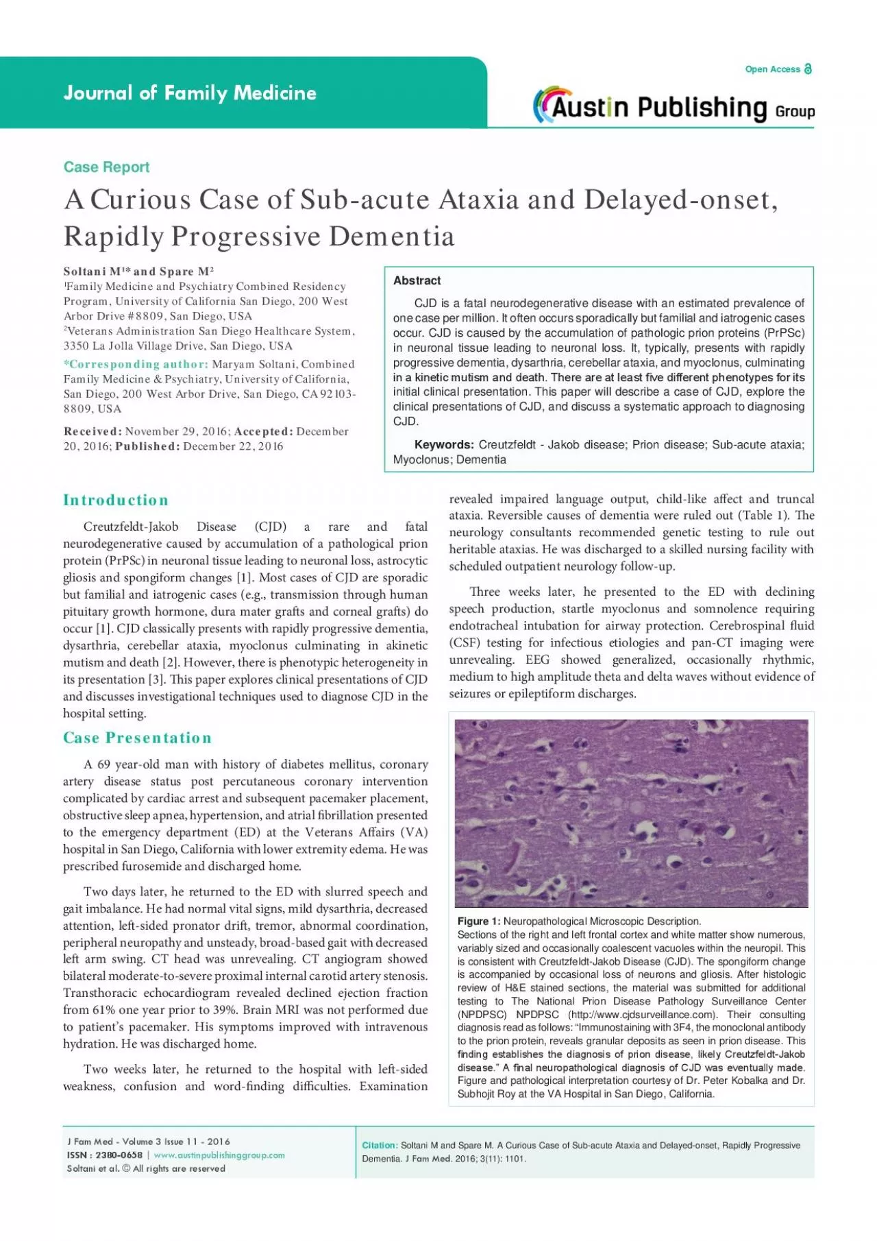
/
revealed impaired language output childlike a31ect and truncal revealed impaired language output childlike a31ect and truncal

revealed impaired language output childlike a31ect and truncal - PDF document

amelia
amelia . @amelia
Follow
342 views
Uploaded On 2022-08-22

revealed impaired language output childlike a31ect and truncal - PPT Presentation

ataxia Reversible causes of dementia were ruled out Table 1 30e neurology consultants recommended genetic testing to rule out heritable ataxias He was discharged to a skilled nursing facility ID: 939682

hospital cjd revealed discharged cjd hospital discharged revealed speech myoclonus unrevealing showed sided gait returned disease neuronal weeks cases

Share:

Link:

Embed:

Download Presentation from below link

Download Pdf The PPT/PDF document "revealed impaired language output childl..." is the property of its rightful owner. Permission is granted to download and print the materials on this web site for personal, non-commercial use only, and to display it on your personal computer provided you do not modify the materials and that you retain all copyright notices contained in the materials. By downloading content from our website, you accept the terms of this agreement.


Presentation Transcript

revealed impaired language output, child-like aect and truncal ataxia. Reversible causes of dementia were ruled out (Table 1). e neurology consultants recommended genetic testing to rule out heritable ataxias. He was discharged to a skilled nursing facility with scheduled outpatient neurology follow-up. ree weeks later, he presented to the ED with declining speech production, startle myoclonus and somnolence requiring endotracheal intubation for airway protection. Cerebrospinal uid (CSF) testing for infectious etiologies and pan-CT imaging were unrevealing. EEG showed generalized, occasionally rhythmic, medium to high amplitude theta and delta waves withou

t evidence of seizures or epileptiform discharges. Introduction Creutzfeldt-Jakob Disease (CJD) a rare and fatal neurodegenerative caused by accumulation of a pathological prion protein (PrPSc) in neuronal tissue leading to neuronal loss, astrocytic gliosis and spongiform changes [1]. Most cases of CJD are sporadic but familial and iatrogenic cases (e.g., transmission through human pituitary growth hormone, dura mater gras and corneal gras) do occur [1]. CJD classically presents with rapidly progressive dementia, dysarthria, cerebellar ataxia, myoclonus culminating in akinetic mutism and death [2]. However, there is phenotypic heterogeneity in its presentation [3].

is paper explores clinical presentations of CJD and discusses investigational techniques used to diagnose CJD in the hospital setting. Case Presentation A 69 year-old man with history of diabetes mellitus, coronary artery disease status post percutaneous coronary intervention complicated by cardiac arrest and subsequent pacemaker placement, obstructive sleep apnea, hypertension, and atrial brillation presented to the emergency department (ED) at the Veterans Aairs (VA) hospital in San Diego, California with lower extremity edema. He was prescribed furosemide and discharged home. Two days later, he returned to the ED with slurred speech and gait imbalance. He had

normal vital signs, mild dysarthria, decreased attention, le-sided pronator dri, tremor, abnormal coordination, peripheral neuropathy and unsteady, broad-based gait with decreased le arm swing. CT head was unrevealing. CT angiogram showed bilateral moderate-to-severe proximal internal carotid artery stenosis. Transthoracic echocardiogram revealed declined ejection fraction from 61% one year prior to 39%. Brain MRI was not performed due to patient’s pacemaker. His symptoms improved with intravenous hydration. He was discharged home. Two weeks later, he returned to the hospital with le-sided weakness, confusion and word-nding diculties. Examinat