/
Diverticula of the Small Intestine Caused by Vitelline Abnormalities; A comparison of Diverticula of the Small Intestine Caused by Vitelline Abnormalities; A comparison of

Diverticula of the Small Intestine Caused by Vitelline Abnormalities; A comparison of - PowerPoint Presentation

anderson
anderson . @anderson
Follow
27 views
Uploaded On 2024-02-09

Diverticula of the Small Intestine Caused by Vitelline Abnormalities; A comparison of - PPT Presentation

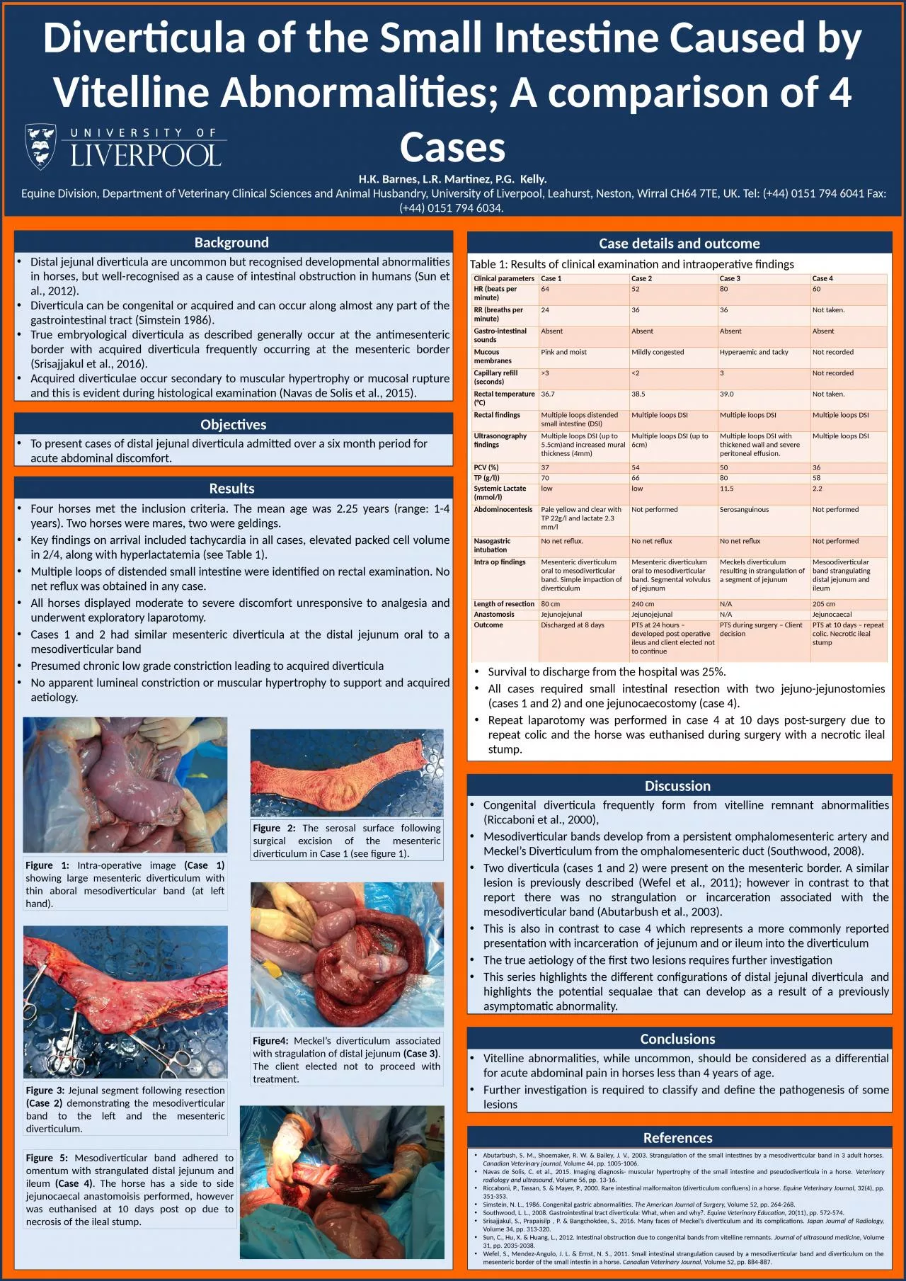
HK Barnes LR Martinez PG Kelly Equine Division Department of Veterinary Clinical Sciences and Animal Husbandry University of Liverpool Leahurst Neston Wirral CH64 7TE UK Tel 44 0151 794 6041 Fax 44 ID: 1045462

diverticulum diverticula mesodiverticular case diverticula diverticulum case mesodiverticular loops band small mesenteric distal volume journal veterinary cases intestinal horses

Share:

Link:

Embed:

Download Presentation from below link

Download Presentation The PPT/PDF document "Diverticula of the Small Intestine Cause..." is the property of its rightful owner. Permission is granted to download and print the materials on this web site for personal, non-commercial use only, and to display it on your personal computer provided you do not modify the materials and that you retain all copyright notices contained in the materials. By downloading content from our website, you accept the terms of this agreement.


Presentation Transcript

1. Diverticula of the Small Intestine Caused by Vitelline Abnormalities; A comparison of 4 CasesH.K. Barnes, L.R. Martinez, P.G. Kelly. Equine Division, Department of Veterinary Clinical Sciences and Animal Husbandry, University of Liverpool, Leahurst, Neston, Wirral CH64 7TE, UK. Tel: (+44) 0151 794 6041 Fax: (+44) 0151 794 6034. Background Distal jejunal diverticula are uncommon but recognised developmental abnormalities in horses, but well-recognised as a cause of intestinal obstruction in humans (Sun et al., 2012). Diverticula can be congenital or acquired and can occur along almost any part of the gastrointestinal tract (Simstein 1986). True embryological diverticula as described generally occur at the antimesenteric border with acquired diverticula frequently occurring at the mesenteric border (Srisajjakul et al., 2016). Acquired diverticulae occur secondary to muscular hypertrophy or mucosal rupture and this is evident during histological examination (Navas de Solis et al., 2015). ObjectivesTo present cases of distal jejunal diverticula admitted over a six month period for acute abdominal discomfort.Results Case details and outcomeConclusions Vitelline abnormalities, while uncommon, should be considered as a differential for acute abdominal pain in horses less than 4 years of age. Further investigation is required to classify and define the pathogenesis of some lesionsReferences Four horses met the inclusion criteria. The mean age was 2.25 years (range: 1-4 years). Two horses were mares, two were geldings. Key findings on arrival included tachycardia in all cases, elevated packed cell volume in 2/4, along with hyperlactatemia (see Table 1).Multiple loops of distended small intestine were identified on rectal examination. No net reflux was obtained in any case. All horses displayed moderate to severe discomfort unresponsive to analgesia and underwent exploratory laparotomy.Cases 1 and 2 had similar mesenteric diverticula at the distal jejunum oral to a mesodiverticular bandPresumed chronic low grade constriction leading to acquired diverticulaNo apparent lumincal constriction or muscular hypertrophy to support and acquired aetiology.Figure 1: Intra-operative image (Case 1) showing large mesenteric diverticulum with thin aboral mesodiverticular band (at left hand). Discussion Congenital diverticula frequently form from vitelline remnant abnormalities (Riccaboni et al., 2000),Mesodiverticular bands develop from a persistent omphalomesenteric artery and Meckel’s Diverticulum from the omphalomesenteric duct (Southwood, 2008).Two diverticula (cases 1 and 2) were present on the mesenteric border. A similar lesion is previously described (Wefel et al., 2011); however in contrast to that report there was no strangulation or incarceration associated with the mesodiverticular band (Abutarbush et al., 2003).This is also in contrast to case 4 which represents a more commonly reported presentation with incarceration of jejunum and or ileum into the diverticulumThe true aetiology of the first two lesions requires further investigationThis series highlights the different configurations of distal jejunal diverticula and highlights the potential sequalae that can develop as a result of a previously asymptomatic abnormality.Figure4: Meckel’s diverticulum associated with stragulation of distal jejunum (Case 3). The client elected not to proceed with treatment.Figure 5: Mesodiverticular band adhered to omentum with strangulated distal jejunum and ileum (Case 4). The horse has a side to side jejunocaecal anastomoisis performed, however was euthanised at 10 days post op due to necrosis of the ileal stump.Figure 3: Jejunal segment following resection (Case 2) demonstrating the mesodiverticular band to the left and the mesenteric diverticulum.Clinical parametersCase 1Case 2Case 3Case 4HR (beats per minute)64528060RR (breaths per minute)243636Not taken.Gastro-intestinal soundsAbsentAbsentAbsentAbsentMucous membranesPink and moistMildly congestedHyperaemic and tackyNot recordedCapillary refill (seconds)>3 <2 3Not recordedRectal temperature (°C)36.738.539.0Not taken.Rectal findingsMultiple loops distended small intestine (DSI)Multiple loops DSIMultiple loops DSIMultiple loops DSIUltrasonography findingsMultiple loops DSI (up to 5.5cm)and increased mural thickness (4mm)Multiple loops DSI (up to 6cm)Multiple loops DSI with thickened wall and severe peritoneal effusion.Multiple loops DSIPCV (%)37545036TP (g/l))70668058Systemic Lactate (mmol/l)lowlow11.52.2AbdominocentesisPale yellow and clear with TP 22g/l and lactate 2.3 mm/lNot performedSerosanguinous Not performedNasogastric intubationNo net reflux.No net refluxNo net reflux Not performedIntra op findingsMesenteric diverticulum oral to mesodiverticular band. Simple impaction of diverticulumMesenteric diverticulum oral to mesodiverticular band. Segmental volvulus of jejunumMeckels diverticulum resulting in strangulation of a segment of jejunumMesoodiverticular band strangulating distal jejunum and ileumLength of resection80 cm240 cmN/A205 cmAnastomosisJejunojejunalJejunojejunalN/AJejunocaecalOutcomeDischarged at 8 daysPTS at 24 hours – developed post operative ileus and client elected not to continuePTS during surgery – Client decisionPTS at 10 days – repeat colic. Necrotic ileal stumpTable 1: Results of clinical examination and intraoperative findingsSurvival to discharge from the hospital was 25%.All cases required small intestinal resection with two jejuno-jejunostomies (cases 1 and 2) and one jejunocaecostomy (case 4). Repeat laparotomy was performed in case 4 at 10 days post-surgery due to repeat colic and the horse was euthanised during surgery with a necrotic ileal stump. Abutarbush, S. M., Shoemaker, R. W. & Bailey, J. V., 2003. Strangulation of the small intestines by a mesodiverticular band in 3 adult horses. Canadian Veterinary journal, Volume 44, pp. 1005-1006.Navas de Solis, C. et al., 2015. Imaging diagnosis- muscular hypertrophy of the small intestine and pseudodiverticula in a horse. Veterinary radiology and ultrasound, Volume 56, pp. 13-16.Riccaboni, P., Tassan, S. & Mayer, P., 2000. Rare intestinal malformaiton (diverticulum confluens) in a horse. Equine Veterinary Journal, 32(4), pp. 351-353.Simstein, N. L., 1986. Congenital gastric abnormalities. The American Journal of Surgery, Volume 52, pp. 264-268.Southwood, L. L., 2008. Gastrointestinal tract diverticula: What, when and why?. Equine Veterinary Education, 20(11), pp. 572-574.Srisajjakul, S., Prapaisilp , P. & Bangchokdee, S., 2016. Many faces of Meckel’s diverticulum and its complications. Japan Journal of Radiology, Volume 34, pp. 313-320.Sun, C., Hu, X. & Huang, L., 2012. Intestinal obstruction due to congenital bands from vitelline remnants. Journal of ultrasound medicine, Volume 31, pp. 2035-2038.Wefel, S., Mendez-Angulo, J. L. & Ernst, N. S., 2011. Small intestinal strangulation caused by a mesodiverticular band and diverticulum on the mesenteric border of the small intestin in a horse. Canadian Veterinary Journal, Volume 52, pp. 884-887.Figure 2: The serosal surface following surgical excision of the mesenteric diverticulum in Case 1 (see figure 1).