/
Development of Skeletal System Development of Skeletal System

Development of Skeletal System - PowerPoint Presentation

anya
anya . @anya
Follow
0 views
Uploaded On 2024-03-13

Development of Skeletal System - PPT Presentation

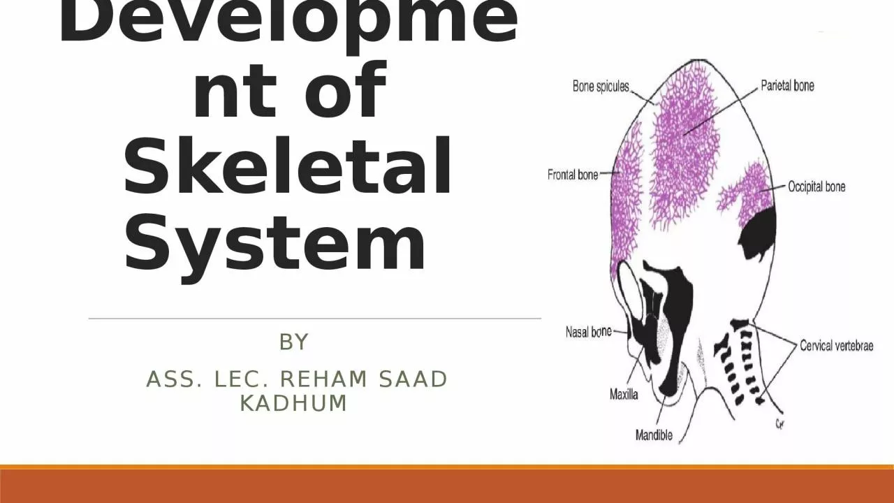
By Ass Lec Reham Saad Kadhum Source of skeletal system In general the skeletal system develops from Mesoderm and Ectoderm paraxial and lateral píate parietal layer ID: 1047032

bone bones skull development bones bone development skull derived flat limb ossification form forms mesoderm week cells cartilaginous part

Share:

Link:

Embed:

Download Presentation from below link

Download Presentation The PPT/PDF document "Development of Skeletal System" is the property of its rightful owner. Permission is granted to download and print the materials on this web site for personal, non-commercial use only, and to display it on your personal computer provided you do not modify the materials and that you retain all copyright notices contained in the materials. By downloading content from our website, you accept the terms of this agreement.


Presentation Transcript

1. Development of Skeletal System By Ass. Lec. Reham Saad Kadhum

2. Source of skeletal system In general, the skeletal system develops from ( Mesoderm and Ectoderm ) paraxial and lateral píate (parietal layer) mesoderm neural crest. ( ectoderm )

3. Ossification Intramembranous ossificationBone formation in which the mesenchyme differentiated directly into the bone e.g. flat bones of the skull.Endochondral ossification The process of bone formation in which the mesenchymal cells give rise to cartilaginous models first which in turn become ossified and form bone e.g. long bones of the limb

4.

5. SkullThe skull can be divided into 2 parts 1. The Neurocranium, (which forms a protective case for the brain).2. The Viscerocranium, (which formed the skeleton of the face).

6. Development of the Skull 1. The Neurocranium, (The development of the brain box)it’s derived from two portions;A. The membranous part, consisting of flat bones, which surrounded the brain as a vault.B. The cartilaginous part, or chondrocrainum , which forms bone of the base of the skull

7. Development of skull A. Membranous portion; Formed by intramembranous ossification. Mesenchymal cells are derived from neural crest and paraxial mesoderm. Cells then encircle the brain and form most of the flat bones of the skullThese flat bones begin as bony spicules radiating from a primary ossification center. After birth, these flat bones enlarged by osteoblastic cells adding bones at the outer surface with osteoclastic activity absorbing the bones from the inner surface.

8. Development of skull A. Membranous portion; At birth, these flat bones are separated by C.T. called the sutures, and larger C.T. called the fontanelles that are derived from the neural crest.This C.T. allows overlapping or called molding of these flat bones to decrease the size of the skull during its passage out of the birth canal. Remodeling of the flat bone occurs after birth resulting in a larger vault in comparison to the face. The connective tissues of the sutures and the fontanelles ossify later on. Before ossification the fontanels indicates the intracranial pressure.

9.

10. B. The cartilaginous part, or chondrocrainumFormed by a combination of mesodermal sclerotome and neural crest cells Cartilage are form around the brain beginning at the notochord. Parachordal cartilage and three occipital sclerotomes fused to form the base of occipital boneWhile the sphenoid and ethmoidal bones are formed from the hypophysial cartilage and the trabeculae cranii.A number of other mesenchymal condensations arise on either side of the median plate. The lesser wing of the sphenoid bone are formed from ala orbitalis. While, caudally the greater wing of the sphenoid are formed from the ala temporalis. Petrous and mastoid parts of the temporal bone are give rise from periotic capsule. All these pieces of bones fuse with each other to form a strong base of the skull, expect for the openings via which the cranial nerves leaves the skull.

11.

12.

13. 2. The ViscerocraniumMainly they develop from the mesoderm of the pharyngeal arches lying on the sides of the pharynx.A. Membranous Viscerocranium: Dorsal portion (the maxillary process) Undergoes intramembranous ossification and gives rise to the maxilla, the zygomatic bone, the squamous temporal bones. Ventral portion (the mandibular process)Contains the Meckel’s cartilageThis region become surrounded by mesenchymal cells that condenses and ossifies by membranous ossification to form the mandible

14.

15. B. Chondral ViscerocraniumDorsal portionForms the malleus and incus (Meckel’s cartilage)Forms the stapes and the styloid process (Reichert’s cartilage)Ventral portion Ossifies and forms the lesser horn and the upper body of the hyoid boneForms the greater horn and lower body of the hyoid bone Note: bones of the base of the skull anterior to the sella turcica are derived from prechordal part of the neural crest, while the more posterior bone is derived from the paraxial chordal mesoderm.

16. Development of the limb bone: The limb buds projects from the ventrolateralbody wall in the 4th week.They are formed of extensions from thesomatic mesoderm of the lateral plate of themesoderm and covered by ectoderm.At the 6th week, the flat plates of the handand foot develop distally. Later on, constrictions appear between the parts of the limb. The tip of the limb bud shows a thickening ofthe ectoderm called the apical ectodermalridge (AER) that induces elongation and formation of the limb buds.Segmental death occurs in the AER results inthe formation of the digits in the hand and foot.

17. Development of the limb bone: The development of the UL precedes that of the LL by about 1-2 days.During the 7th week, the UL rotate 90 degrees ,laterally, and the LL rotates 90 degreesmedially.In the 6th week, the mesenchyme of the limb buds forms the hyaline cartilaginous model ofthe long bones, which ossify in the 12th week. During this time, primary ossification centers will form the diaphysis of the shaft of the bones.After birth secondary ossification centers appear at the end sides of the shaft of the bones forming the epiphysis.This epiphyseal plate form a cartilaginous rim between the bone of the epiphysis and thediaphysis.The growth of this epiphyseal plate elongates the limb bones.

18.

19. Development of the vertebral column:vertebrae are derived from the sclerotomes of the somites that migrate medially and ventrally to surround the notochord and the spinal cord during the 4th week.The sclerotomes of each of the somite are separated from each other by the intersegmental branches of the dorsal aorta of the embryo. Later on, the caudal part of each of the sclerotome invades the cephalic part of the next sclerotome across the region of the intersegmental vessels between them.

20. Development of the vertebral column:This invasion results in the formation of the cartilaginous body of the vertebrae. The mid-region between the cephalic and caudal parts of each sclerotome forms the intervertebral disc (IVD).The notochord forms the Centrum of the vertebral body and the centrally located nucleus pulposus of the IVD.The peripheral annulus fibrosis of the IVD is derived from the medial regions of the sclerotomes.

21. Development of the Ribs The ribs develop as processed from the thoracic vertebrae that are called as the costal processes.The rib therefore is derived from the thoracic sclerotomes.The sternum is derived from the somatic layer of lateral plate of the mesoderm forming the ventral body wall.

22.

23.