/
Mitochondria Membrane bound cell organelles, associated with cell respiration, the source Mitochondria Membrane bound cell organelles, associated with cell respiration, the source

Mitochondria Membrane bound cell organelles, associated with cell respiration, the source - PowerPoint Presentation

anya
anya . @anya
Follow
64 views
Uploaded On 2024-01-03

Mitochondria Membrane bound cell organelles, associated with cell respiration, the source - PPT Presentation

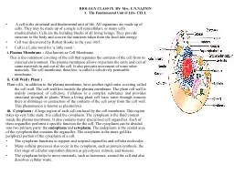
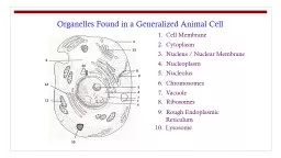
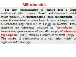
Discovered by Benda 1898 Present in eukaryotic cell Bean shaped organelles 110 um long and about 05um wide occur free in cytoplasm 2 STRUCTURE Bounded by two membrane outer and inner membrane Both membrane are about 6070 A thick ID: 1037464

membrane mitochondrial dna mitochondria mitochondrial membrane mitochondria dna proteins genomes space complex sequences transfer matrix genetic protein translocation intermembrane

Share:

Link:

Embed:

Download Presentation from below link

Download Presentation The PPT/PDF document "Mitochondria Membrane bound cell organel..." is the property of its rightful owner. Permission is granted to download and print the materials on this web site for personal, non-commercial use only, and to display it on your personal computer provided you do not modify the materials and that you retain all copyright notices contained in the materials. By downloading content from our website, you accept the terms of this agreement.


Presentation Transcript

1. Mitochondria

2. Membrane bound cell organelles, associated with cell respiration, the source of energy, being termed as power house of the cellDiscovered by Benda (1898)Present in eukaryotic cellBean shaped organelles, 1-10 um long and about 0.5um wide, occur free in cytoplasm2

3. STRUCTURE: Bounded by two membrane- outer and inner membrane, Both membrane are about 60-70 A thickOuter membrane contain more phospholipid and cholestrol than inner membrane the inner mitochondrial membrane contains an unusually high percentage (greater than 70%) of proteins, which areinvolved in oxidative phosphorylation as well as in the transport of metabolites (e.g., pyruvate and fatty acids) between the cytosol and mitochondria.Space between membrane- perimitochondrial space or inter membrane spaceSpace bounded by inner membrane – inner membrane space, its filled with matrix3

4. Matrix: dense granules, DNA, ribosomes, and enzymes (krebs cycle)Outer membrane is smooth while inner membrane is thrown up into series of folds, called CristaeSeveral thousand particles are present on inner side of the inner membrane – elementary particles or oxysomes or F0-F1 particel4

5. Structure5

6. 6

7. 7

8. 8

9. 9

10. 10

11. 11

12. 12

13. 13

14. Mitochondrial Origin14

15. Mitochondrial Division15

16. 16

17. Semi autonomy:Presence of mtDNA (circular)DNA dependent RNA synthesisPresence of ribosome (70s)Protein synthesisCan multiply by division Contain gene for male sterility 17

18. Mitochondria contain their own genetic system, which is separate and distinct from the nuclear genome of the cell. Mitochondria are thought to have evolved from bacteria that developed a symbiotic relationship in which they lived within larger cells (endosymbiosis). This hypothesis has recently been substantiated by the results of DNA sequence analysis, which revealed striking similarities between the genomes of mitochondria and of the bacterium Rickettsia prowazekii. Rickettsia are intracellular parasites which, like mitochondria, are only able to reproduce within eukaryotic cells. Consistent with their similar symbiotic lifestyles, the genomic DNA sequences of Rickettsia and mitochondria suggest that they share a common ancestor, from which the genetic system of present‐day mitochondria evolved.18

19. Mitochondrial genomes are usually circular DNA molecules, like those of bacteria, which are present in multiple copies per organelle. They vary considerably in size betweendifferent species. The genomes of human and most other animal mitochondria are only about 16 kb, but substantially larger mitochondrial genomes are found in yeasts(approximately 80 kb) and plants (more than 200 kb). However, these larger mitochondrial genomes are composed predominantly of noncoding sequences and do not appear tocontain significantly more genetic information. For example, the largest sequenced mitochondrial genome is that of the plant Arabidopsis thaliana. Although Arabidopsismitochondrial DNA is approximately 367 kb, it encodes only 32 proteins: just more than twice the number encoded by human mitochondrial DNA. Mitochondrial genomes encodeall of the ribosomal RNAs and most of the transfer RNAs needed for translation of these protein‐coding sequences within mitochondria. Other mitochondrial proteins areencoded by nuclear genes, which are thought to have been transferred to the nucleus from the ancestral mitochondrial genome.19

20. 20

21. Mitochondrial GenomeGenerally, mitochondrial DNA encodes:tRNAsrRNAsProtein subunits, including:Polypeptides of cytochrome oxidaseNADH dehydrogenaseATPase21

22. Map of the genes of human mitochondrial DNA22

23. 23

24. 24

25. Exception in Genetic code25

26. 26

27. 27

28. 28

29. Like the DNA of nuclear genomes, mitochondrial DNA can be altered by mutations, which are frequently deleterious to the organelle. Since almost all the mitochondria of fertilized eggs are contributed by the oocyte rather than by the sperm, germ‐line mutations in mitochondrial DNA are transmitted to the next generation by the mother. Such mutations havebeen associated with a number of diseases. For example, Leber's hereditary optic neuropathy, a disease that leads to blindness, can be caused by mutations in mitochondrial genes that encode components of the electron transport chain.Mutation in one of the mitochondrial transfer RNA genes, characterized by a decrease in synthesis of mitochondrial protein required for electron transport chain and ATP synthesis causes a disorder of muscles termed as a. Myoclonic epilepsy and ragged red fiber disease (MERRF).29

30. Import of proteins into mitochondriaProteins are targeted for mitochondria by an amino-terminal presequence containing positively charged amino acids. Proteins are maintained in a partially unfolded state by association with a cytosolic Hsp70 and are recognized by a receptor on the surface of mitochondria. The unfolded polypeptide chains are then translocated through the Tom complex in the outer membrane and transferred to the Tim complex in the inner membrane. The voltage component of the electrochemical gradient is required for translocation across the inner membrane. The presequence is cleaved by a matrix protease, and a mitochondrial Hsp70 binds the polypeptide chain as it crosses the inner membrane, driving further protein translocation. A mitochondrial Hsp60 then facilitates folding of the imported polypeptide within the matrix.30

31. Insertion of mitochondrial membrane proteinsProteins targeted for the mitochondrial membranes contain hydrophobic stop-transfer sequences that halt their translocation through the Tom or Tim complexes and lead to their incorporation into the outer or inner membranes, respectively.31

32. Proteins can be targeted to the intermembrane space by several mechanisms. Some proteins (I) are translocated through the Tom complex and released into the intermembrane space. Other proteins (II) are transferred from the Tom complex to the Tim complex, but they contain hydrophobic stop-transfer sequences that halt translocation through the Tim complex. These stop-transfer sequences are then cleaved to release the proteins into the intermembrane space. Still other proteins (III) are imported to the matrix, as depicted in Figure 10.4. Removal of the presequence within the matrix then exposes a hydrophobic signal sequence, which targets the protein back across the inner membrane to the intermembrane space.32