/
Principles of Principles of

Principles of - PDF document

ariel
ariel . @ariel
Follow
342 views
Uploaded On 2022-09-09

Principles of - PPT Presentation

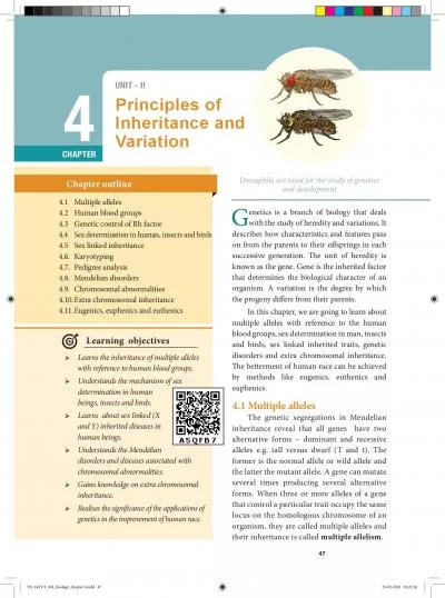
47 UNIT II Inheritance and Variation CHAPTER 4 Multiple alleles Human blood groups Genetic control of Rh factor 44 Sex determination in human insects and birds Sex linked inheritance Karyotypin ID: 953692

146 sex inheritance chromosome sex 146 chromosome inheritance blood chromosomes male human 145 genes gene female determination type normal

Share:

Link:

Embed:

Download Presentation from below link

Download Pdf The PPT/PDF document "Principles of" is the property of its rightful owner. Permission is granted to download and print the materials on this web site for personal, non-commercial use only, and to display it on your personal computer provided you do not modify the materials and that you retain all copyright notices contained in the materials. By downloading content from our website, you accept the terms of this agreement.


Presentation Transcript

47 UNIT - II Principles of Inheritance and Variation CHAPTER 4 Multiple alleles Human blood groups Genetic control of Rh factor 4.4 Sex determination in human, insects and birds Sex linked inheritance Karyotyping Pedigree analysis Mendelian disorders Chromosomal abnormalities Extra chromosomal inheritance Eugenics, euphenics and euthenics Chapter outline Learning objectives Learns the inheritance of multiple alleles with reference to human blood groups. Understands the mechanism of sex determination in human beings, insects and birds. Learns about sex linked (X and Y) inherited diseases in human beings. Understands the Mendelian disorders and diseases associated with chromosomal abnormalities. Gains knowledge on extra chromosomal inheritance.  Realises the significance of the applications of genetics in the improvement of human race. G enetics is a branch of biology that deals with the study of heredity and variations. It describes how characteristics and features pass on from the parents to their osprings in each successive generation. e unit of heredity is known as the gene. Gene is the inherited factor that determines the biological character of an organism. A variation is the degree by which the progeny diers from their parents. In this chapter, we are going to learn about multiple alleles with reference to the human blood groups, sex determination in man, insects and birds, sex linked inherited traits, genetic disorders and extra chromosomal inheritance. e betterment of human race can be achieved by methods like eugenics, euthenics and euphenics. 4.1 Multiple alleles The genetic segregations in Mendelian inheritance reveal that all genes have two alternative forms – dominant and recessive alleles e.g. tall versus dwarf (T and t). The former is the normal allele or wild allele and the latter the mutant allele. A gene can mutate several times producing several alternative forms. When three or more alleles of a gene that control a particular trait occupy the same locus on the homologous chromosome of an organism, they are called multiple alleles and their inheritance is called multiple allelism . Drosophila are ideal for the study of genetics anddevelopment TN_GOVT_XII_Zoology_chapter 4.indd 47 25-02-2019 18:21:56 48 Principles Of Inheritance And Variation determines B antigen and I O allele specifies no antigen. Individuals who possess these antigens in their fluids such as the saliva are called s

ecretors. Each allele (I A and I B ) produces a transferase enzyme. I A allele produces N-acetyl galactose transferase and can add N-acetyl galactosamine (NAG) and I B allele encodes for the enzyme galactose transferase that adds galactose to the precursor (i.e. H substances) In the case of I O /I O allele no terminal transferase enzyme is produced and therefore called “null” allele and hence cannot add NAG or galactose to the precursor. From the phenotypic combinations it is evident that the alleles I A and I B are dominant to I O , but co-dominant to each other (I A =I B ). Their dominance hierarchy can be given as (I A =I B � I O ). A child receives one of three alleles from each parent, giving rise to six possible genotypes and four possible blood types (phenotypes). The genotypes are I A I A , I A I O , I B I B , I B I O , I A I B and I O I O . • Antigens similar to those found among human beings have been recognized in the blood of other organisms. A-type antigens have been found in chimpanzees and in gibbons, A, B and AB antigen in orangutans. • New world monkeys (Platyrrhina) and lemurs have a substance similar but not identical with B antigen in humans. • ree blood groups have been distinguished in cats with a genetic system similar to those in humans. • e secretors (antigens found in the body uids) can be detected in tears, saliva, urine, semen, gastric juice and in the milk of animals. 4.2 Human Blood Groups Multiple allelism occurs in humans, particularly in the inheritance of dierent types of blood groups. e blood group inheritance in human can be understood by learning about antigens and antibodies. e composition of blood, dierent types of blood groups (ABO) the blood antigens and antibodies were discussed in chapter 7 of class XI. 4.2.1 ABO blood types Multiple allele inheritance of ABO blood groups Blood differs chemically from person to person. When two different incompatible blood types are mixed, agglutination (clumping together) of erythrocytes (RBC) occurs. The basis of these chemical differences is due to the presence of antigens (surface antigens) on the membrane of RBC and epithelial cells. Karl Landsteiner discovered two kinds of antigens called antigen ‘A’ and antigen ‘B’ on the surface of RBC’s of human blood. Based on the presence or absence of these antigens three kinds of blood groups, type ‘A

6;, type ‘B’, and type ‘O’ (universal donor)were recognized. The fourth and the rarest blood group ‘AB’ (universal recipient) was discovered in 1902 by two of Landsteiner’s students Von De Castelle and Sturli. Bernstein in 1925 discovered that the inheritance of different blood groups in human beings is determined by a number of multiple allelic series. The three autosomal alleles located on chromosome 9 are concerned with the determination of blood group in any person. The gene controlling blood type has been labeled as ‘L’ (after the name of the discoverer, Landsteiner) or I (from isoagglutination). The I gene exists in three allelic forms, I A , I B and I O . I A specifies A antigen. I B allele TN_GOVT_XII_Zoology_chapter 4.indd 48 25-02-2019 18:21:57 Principles Of Inheritance And VariationTable 4.1 Genetic basis of the human ABO blood groups GenotypeABO blood group phenotype Antigens present on red blood cellAntibodies present in blood plasmaType AAnti -BType AAnti -BType BAnti -AType BAnti -AType ABA and BNeither Anti -A norAnti-BType ONeither A nor BAnti -A and anti -BRhesus or Rh – FactorThe Rh factor or Rh antigen is found on the surface of erythrocytes. It was discovered in 1940 by Karl Landsteiner and Alexander Wiener in the blood of rhesus monkey, Macacarhesus and later in human beings. The term ‘Rh factor’ refers to “immunogenic D antigen of the Rh blood group system. An individual having D antigen are Rh D positive (Rh) and those without D antigen are Rh D negative (Rh)”. Rhesus factor in the blood is inherited as a dominant trait. Naturally occurring Anti D antibodies are absent in the plasma of any normal individual. However if an Rh (Rh negative) person is exposed to Rh(Rh positive) blood cells (erythrocytes) for the first time, anti D antibodies are formed in the blood of that individual. On the other hand, when an Rh positive person receives Rh negative blood no effect is seen. 4.3 Genetic control of Rh factorFisher and Race hypothesis:Rh factor involves three dierent pairs of alleles located on three dierent closely linked loci on the chromosome pair. is system is more commonly in use today, and uses the 'Cde' nomenclature. C or c C or cD or dD or dE or eE or eFig. 4.1 Fischer and Race hypothesis – Rh Blood Type - Homologous Chromosome pair (showing 3 loci and 2 alleles per locus)In the above Fig. 4.1, three pairs of Rh alleles (Cc, Dd and Ee) occur at 3 d

ierent loci on homologous chromosome pair-1. e possible genotypes will be one C or c, one D or d, one E or e from each chromosome. For e.g. CDE/cde; CdE/cDe; cde/cde; CDe/CdE etc., All genotypes carrying a dominant ‘D’ allele will produce Rhpositive phenotype and double recessive genotype ‘dd’ will give rise to Rhnegative phenotype.Wiener HypothesisWiener proposed the existence of eight alleles (R, r, r) at a single Rh locus. All genotypes carrying a dominant ‘R allele’ (R) will produce Rhpositive’ phenotype and double recessive genotypes (rr, rr, rr, rr) will give rise to Rhnegative phenotype.4.3.1 Incompatibility of Rh Factor – Erythroblastosis foetalisRh incompatability has great signicance in child birth. If a woman is Rh negative and TN_GOVT_XII_Zoology_chapter 4.indd 49 25-02-2019 18:21:57 50 Principles Of Inheritance And Variationthe man is Rh positive, the foetus may be Rh positive having inherited the factor from its father. e Rh negative mother becomes sensitized by carrying Rh positive foetus within her body. Due to damage of blood vessels, during child birth, the mother’s immune system recognizes the Rh antigens and gets sensitized. e sensitized mother produces Rh antibodies. e antibodies are IgG type which are small and can cross placenta and enter the foetal circulation. By the time the mother gets sensitized and produce anti ‘D’ antibodies, the child is delivered.Usually no eects are associated with exposure of the mother to Rh positive antigen during the rst child birth, subsequent Rh positive children carried by the same mother, may be exposed to antibodies produced by the mother against Rh antigen, which are carried across the placenta into the foetal blood circulation. is causes haemolysis of foetal RBC resulting in haemolytic jaundice and anaemia. is condition is known as Erythoblastosis foetalis or Haemolytic disease of the new born (HDN).Prevention of Eryhroblastosis foetalisIf the mother is Rh negative and foetus is Rh positive, anti D antibodies should be administered to the mother at 28 and 34week of gestation as a prophylactic measure. If the Rh negative mother delivers Rh positive child then anti D antibodies should be administered to the mother soon aer delivery. is develops passive immunity and prevents the formation of anti D antibodies in the mothers blood by destroying the Rh foetal RBC before the mother’s immune system is sensitized

. is has to be done whenever the woman attains pregnancy.4.4 Sex DeterminationSex determination is the method by which the distinction between male and female is established in a species. Sex chromosomes determine the sex of the individual in dioecious or unisexual organisms. The chromosomes other than the sex chromosomes of an individual are called autosomes. Sex chromosomes may be similar (homomorphic) in one sex and dissimilar (heteromorphic) in the other. Individuals having homomorphic sex chromosomes produce only one type of gametes (homogametic) whereas heteromorphic individuals produce two types of gametes (heterogametic).Chromosomal basis of sex determinationHeterogametic Sex Determination:In heterogametic sex determination one of the sexes produces similar gametes and the other sex produces dissimilar gametes. Thesex of the offspring is determined at the time of fertilization.Heterogametic MalesIn this method of sex determination the males are heterogametic producing dissimilar gametes while females are homogametic producing similar gametes. It is of two kinds XX-XO type and XX-XY type.XXXO TypeThis method of sex determination is seenin bugs, some insects such as cockroaches and grasshoppers. The female with two X chromosomes are homogametic (XX) while the males with only one X chromosome Y CHROMOSOME e human Y chromosome is only 60 Mb in size with 60 functional genes whereas X chromosomes are 165 Mb in size with about 1,000 genes. TN_GOVT_XII_Zoology_chapter 4.indd 50 25-02-2019 18:21:57 Principles Of Inheritance And Variation 51 are heterogametic (XO). The presence of an unpaired X chromosomes determines the male sex. The males with unpaired ‘X’ chromosome produce two types of sperms, one half with X chromosome and other half without X chromosome. The sex of the offspring depends upon the sperm that fertilizes the egg (Fig. 4.2) . P 1 AAXXAAXO Gametes AX AX AO F1 Generation AAXXAAXO Fig. 4.2 XX-XO Type of sex determination XXXY type (Lygaeus Type) This method of sex determination is seen in human beings and in Drosophila . The females are homogametic with XX chromosome, while the males are heterogametic with X and Y chromosome. Homogametic females produce only one kind of egg, each with one X chromosome, while the heterogametic males produce two kinds of sperms some with X chromosome and some with Y chromosome. The sex of the embryo depends on the fertilizing sperm. An egg fertilized by an ‘X&

#146; bearing sperm produces a female, if fertilized by a ‘Y’ bearing sperm, a male is produced (Fig. 4.3). P 1 AAXXAAXY Gametes AX AX AY F1 Generation AAXXAAXY Fig. 4.3 XX-XY Type of sex determination Heterogametic Females In this method of sex determination, the homogametic male possesses two ‘X’ chromosomes as in certain insects and certain vertebrates like fishes, reptiles and birds producing a single type of gamete; while females produce dissimilar gametes. The female sex consists of a single ‘X’ chromosome or one ‘X’ and one ‘Y’ chromosome. Thus the females are heterogametic and produce two types of eggs. To avoid confusion with the XX-XO and XX-XY types of sex determination, the alphabets ‘Z’ and ‘W’ are used here instead of X and Y respectively. Heterogametic females are of two types, ZO-ZZ type and ZW-ZZ type. ZOZZ Type This method of sex determination is seen in certain moths, butterflies and domestic chickens. In this type, the female possesses single ‘Z’ chromosome in its body cells and is heterogametic (ZO) producing two kinds of eggs some with ‘Z’ chromosome and some without ‘Z’ chromosome, while the male possesses two ‘Z’ chromosomes and is homogametic (ZZ) (Fig. 4.4). P 1 AAZO AAZZ Gametes AZ AO AZ F1 Generation AAZZ Fig. 4.4 ZO-ZZ type of sex determination ZW-ZZ type is method of sex determination occurs in certain insects (gypsy moth) and in vertebrates such as shes, reptiles and birds. In this method the female has one ‘Z’ and one ‘W’ chromosome (ZW) producing two types of eggs, some carrying the Z chromosomes and some carry the W chromosome. e male sex TN_GOVT_XII_Zoology_chapter 4.indd 51 25-02-2019 18:21:57 52 Principles Of Inheritance And Variationhas two ‘Z’ chromosomes and is homogametic (ZZ) producing a single type of sperm (Fig .4.5). P 1 AAZW AAZZ Gametes AZ AW AZ F1 Generation AAZZ AAZW Fig. 4.5 ZW-ZZ type of sex determinationSex determintion in human beingsGenes determining sex in human beings are located on two sex chromosomes, called allosomes. In mammals, sex determination is associated with chromosomal dierences between the two sexes, typically XX females and XY males. 23 pairs of human chromosomes include 22 pairs of autosomes (44A) and one pair of sex chromosomes (XX or XY). Females are homogametic producin

g only one type of gametes (egg), each containing one X chromosome while the males are heterogametic producing two types of sperms with X and Y chromosomes. An independently evolved XX: XY system of sex chromosomes also exist in Drosophila(Fig. 4.6)Fig. 4.6 Sex determination in human beingse Y Chromosome and Male DevelopmentCurrent analysis of Y chromosomes has revealed numerous genes and regions with Male eterogametic) (Homogametic) Parents 44A + XY 4A + XX Gametes Sperms Ova (22A+X) (22A+Y) (22A+X) (22A+X) Osprings/ Progeny ( 44A+XX 44A+XY +XX 44A+XY emale Male emale Male potential genetic function; some genes with or without homologous counterparts are seen on the X. Present at both ends of the Y chromosome are the pseudoautosomal regions (PARs) that are similar with regions on the X chromosome which synapse and recombine during meiosis. e remaining 95% of the Y chromosome is referred as the Non - combining Region of the Y (NRY). e NRY is divided equally into functional genes (euchromatic) and non functional genes (heterochromatic). Within the euchromatin regions, is a gene called Sex determining region Y (SRY). In humans, absence of Y chromosome inevitably leads to female development and this SRY gene is absent in X chromosome. e gene product of SRY is the testes determining factor (TDF) present in the adult male testis.4.4.1 Genic balance in DrosophilaGenic balance mechanisms of sex determination in Drosophila was first studied by C.B. Bridges. In Drosophila, the presence of Y chromosome is essential for the fertility of male sex, but does not determine the male sex. The gene for femaleness is located on the X chromosome and those for maleness are located on the autosomes. When geneticist C.B. Bridges, working with Drosophila, crosseda triploid (3n) female with a normal male, he observed many combinations of autosomes and sex chromosomes in the offspring. From his results Bridges in 1921 suggested that sex Drosophila is determined by the balance between the genes for femaleness located on the ‘X’ chromosomes and those for maleness located on the ‘autosomes’ . Hence the sex of an individual is determined by the ratio of its X chromosome to that of its autosome sets. This ratio is termed sexindex and is expressed as: Number of X ChromosomesNumber of Sets of Autosomes Sex index = TN_GOVT_XII_Zoology_chapter 4.indd 52 25-02-2019 18:21:57 Principles Of Inheritance And

VariationChange in this ratio leads to a changed sex phenotype. e results obtained from a cross between triploid female Drosophila (3A:3X) with a diploid male (2A: XY) is shown in tables 4.2. and 4.3.Table: 4.2 Bridges classical cross of a triploid (3A+XXX) female y and a diploid (2A+XY) male y Triploid Diploid Parent XX 2A + XY Gametes + XX) (A + X) (A + X) (A + Y) (2A + X) (A + XX) 2A+XX3A + XXXTriploid Female3A + XXYTriploid Intersex3A + XXTriploid Intersex3A + XYSuper MaleA+XX2A + XXXSuper female2A + XXYDiploid Female2A + XXDiploid Female2A + XYDiploid MaleWhenthe A ratio is 1.00 as in a normal female, or greater than 1.00, the organism is a female. When this ratio is 0.50 as in a normal male or less than 0.50 the organism is a male. At 0.67, the organism is an intersex. metamales (X/A = 0.33) and metafemales (X/A=1.50) are usually very weak and sterile.A sex–switch gene in Drosophila directs female development. is gene, sex–lethal (SxL) located on the X chromosome, has two states of activity. When it is ‘on’ it directs female development and when it is ‘o’ maleness ensures. Other genes located on the X chromosome and autosomes regulate this sex-switch gene. However, the Y- chromosome of Drosophila is required for male fertility.Table: 4.3 Dierent doses of X chromosomes and autosome sets and their eect on sex determination in Drosophila PhenotypeNumber of ‘X’ Chromosomes (X)Number of Autosome sets Number of X chromosomeSex Index = Number of autosome setsMeta female / Super female 3/2 = Normal FemaleTetraploid 4/4 = Triploid 3/3 = Diploid 2/2 = Haploid 1/1 = Inter sex 2/3 = Normal male ½ = Meta male / Super male 1/3 = 0.33 • X-Chromosome was discovered byHenking (1891) • Y-Chromosome was discovered byStevens (1902) TN_GOVT_XII_Zoology_chapter 4.indd 53 25-02-2019 18:21:57 54 Principles Of Inheritance And VariationGynandromorphsese individuals have parts of their body expressing male characters and other parts of the body expressing female characters. e organism is made up of tissues of male and female genotype and represents a mosaic pattern.4.4.2 Dosage compensation - Barr bodyIn 1949, Barr and Bertram rst observed a condensed body in the nerve cells of female cat which was absent in the male. is condensed body was called sex chromatin by them and was later referred as Barr body. In the XY chromosomal system of sex determina

tion, males have only one X chromosome, whereas females have two. A question arises: how does the organism compensate for this dosage dierences between the sexes? In mammals the necessary dosage compensation is accomplished by the inactivation of one of the X chromosome in females so that both males and females have only one functional X chromosome per cell.Mary Lyon suggested that Barr bodies represented an inactive chromosome, which in females becomes tightly coiled into a heterochromatin, a condensed and visible form of chromatin (Lyon’s hypothesis). e number of Barr bodies observed in cell was one less than the number of X-Chromosome. XO females have no Barr body, whereas XXY males have one Barr body. • The number of Barr bodies followsN-1 rule (N minus one rule), where Nis the total number of X chromosomespresent.Haplodiploidy in Honeybees n hymenopteran insects such as honeybees, ants and wasps a mechanism of sex determination called haplodiploidy mechanism of sex determination is common. In this system, the sex of the ospring is determined by the number of sets of chromosomes it receives. Fertilized eggs develop into females (Queen or Worker) and unfertilized eggs develop into males (drones) by parthenogenesis. It means that the males have half the number of chromosomes (haploid) and the females have double the number (diploid), hence the name haplodiplody for this system of sex determination.is mode of sex determination facilitates the evolution of sociality in which only one diploid female becomes a queen and lays the eggs for the colony. All other females which are diploid having developed from fertilized eggs help to raise the queen’s eggs and so contribute to the queen’s reproductive success and indirectly to their own, a phenomenon known as Kin Selection. e queen constructs their social environment by releasing a hormone that suppresses fertility of the workers.4.5 Sex Linked Inheritancee inheritance of a trait that is determined by a gene located on one of the sex chromosomes is called sex linked inheritance. Genes present on the dierential region of X or Y chromosomes are called sex linked genes. e genes present in the dierential region of “X” chromosome are called X linked genes. e X–linked genes have no corresponding alleles in the Y chromosome. e genes present in the dierential region of Y chromosome are called Y- linked or holandric genes. e Y linked genes have no cor

responding allele in X chromosome. e Y linked genes inherit along with Y chromosome and they phenotypically express only in the male sex. Sex linked inherited traits are more common in males than females because, males are hemizygous and therefore express the trait when they inherit one mutant allele. TN_GOVT_XII_Zoology_chapter 4.indd 54 25-02-2019 18:21:57 Principles Of Inheritance And Variatione X – linked and Y – linked genes in the dierential region (non–homologus region) do not undergo pairing or crossing over during meiosis. e inheritance of X or Y linked genes is called sex-linked inheritance.4.5.1 Inheritance of X - linked genesRed-green colour blindness or daltonism, haemophilia and Duchenne’s muscular dystrophy are examples of X-linked gene inheritance in humans.1.HaemophiliaHaemophilia is commonly known asbleeder’s disease, which is more common in men than women. is hereditary disease was rst reported by John Cotto in 1803. Haemophilia is caused by a recessive X-linked gene. A person with a recessive gene for haemophilia lacks a normal clotting substance (thromboplastin) in blood, hence minor injuries cause continuous bleeding, leading to death. e females are carriers of the disease and would transmit the disease to 50% of their sons even if the male parent is normal. Haemophilia follows the characteristic criss - cross pattern of inheritance.2.Colour blindnessIn human beings a dominant X – linkedgene is necessary for the formation of colour sensitive cells, the cones. e recessive form of this gene is incapable of producing colour sensitive cone cells. Homozygous recessive females (X) and hemizygous recessive males Y) are unable to distinguish red and green colour. e inheritance of colour blindness can be studied in the following two types of marriages. (i) arriage between colour blind man and normal visioned womanA marriage between a colour blind manand a normal visioned woman will produce normal visioned male and female individuals generation but the females are carriersThe marriage between a F normal visioned carrier woman and a normal visioned male will produce one normal visioned female, one carrier female, one normal visioned male and one colour blind male. Colour blind trait is inherited from the male parent to his grandson through carrier daughter, which is an example of criss-cross pattern of inheritance (Fig. 4.7). Fig. 4.7 Marriage between colour blind man and normal visioned wom

an ii) arriage between normal visioned man and colour blind womanIf a colour blind woman (X) marries a normal visioned male (XY), all Fsons will be colourblind and daughters will be normal visioned but are carriers. Marriage between F carrier female with a colour blind male will produce normal visioned carrier daughter, colourblind daughter, normal visioned son and a colourblind son in the generation (Fig. 4.8). TN_GOVT_XII_Zoology_chapter 4.indd 55 25-02-2019 18:21:57 56 Principles Of Inheritance And Variation &RnRXUEnLQGIHoDnH Fig. 4.8 Marriage between normal visioned man and colour blind woman4.5.2 Inheritance of Y- linked genesGenes in the non-homologous region of the Y-chromosome are inherited directly from male to male. In humans, the Y-linked or holandric genes for hypertrichosis (excessive development of hairs on pinna of the ear) are transmitted directly from father to son, because males inherit the Y chromosome from the father. Female inherits only X chromosome from the father and are not aected.4.6 KaryotypingKaryotyping is a technique through which a complete set of chromosomes is separated from a cell and the chromosomes are arranged in pairs. An idiogram refers to a diagrammatic representation of chromosomes.Preparation of KaryotypeTjio and Levan (1960) described a simple method of culturing lymphocytes from the human blood. Mitosis is induced followed by addition of colchicine to arrest cell division at metaphase stage and the suitable spread of metaphase chromosomes is photographed. e individual chromosomes are cut from the photograph and are arranged in an orderly fashion in homologous pairs. is arrangement is called a karyotype. Chromosome banding permits structural denitions and dierentiation of chromosomes.Applications of Karyotyping: • It helps in gender identification. • It is used to detect the chromosomalaberrations like deletion, duplication,translocation, nondisjunction ofchromosomes. • It helps to identify the abnormalities ofchromosomes like aneuploidy. • It is also used in predicting the evolutionaryrelationships between species. • Genetic diseases in human beings can bedetected by this technique.Human KaryotypeDepending upon the position of the centromere and relative length of two arms, human chromosomes are of three types: Metacentric, sub metacentric and acrocentric. The photograph of chromosomes are arranged in the order of descending length in groups from A to G (Fig. 4.9). &

#x0014;  Fig. 4.9 - Human karyotype (male) TN_GOVT_XII_Zoology_chapter 4.indd 56 25-02-2019 18:21:57 Principles Of Inheritance And Variation 57 4.7 Pedigree Analysis Pedigree is a “family tree”, drawn with standard genetic symbols, showing the inheritance pathway for specic phenotypic characters. (Fig. 4.10). Pedigree analysis is the study of traits as they have appeared in a given family line for several past generations. a single gene to the addition or subtraction of an entire chromosome or even a set of chromosomes. Genetic disorders are of two types namely, Mendelian disorders and chromosomal disorders. 4.8 Mendelian disorders Alteration or mutation in a single gene causes Mendelian disorders. ese disorders are transmitted to the osprings on the same line as the Mendelian pattern of inheritance. Some examples for Mendelian disorders are alassemia, albinism, phenylketonuria, sickle cell anaemia, Huntington's chorea, etc., ese disorders may be dominant or recessive and autosomal or sex linked. alassemia alassemia is an autosomal recessive disorder. It is caused by gene mutation resulting in excessive destruction of RBC’s due to the formation of abnormal haemoglobin molecules. Normally haemoglobin is composed of four polypeptide chains, two alpha and two beta globin chains. alassemia patients have defects in either the alpha or beta globin chain causing the production of abnormal haemoglobin molecules resulting in anaemia. alassemia is classied into alpha and beta based on which chain of haemoglobin molecule is aected. It is controlled by two closely linked Genetic Disorders A genetic disorder is a disease or syndrome that is caused by an abnormality in an individual DNA. Abnormalities can range from a small mutation in Fig. 4.10 Symbols commonly used in pedigree charts TN_GOVT_XII_Zoology_chapter 4.indd 57 25-02-2019 18:21:58 58 Principles Of Inheritance And VariationHuntington’s choreaIt is inherited as an autosomal dominant lethal gene in man. It is characterized by involuntary jerking of the body and progressive degeneration of the nervous system, accompanied by gradual mental and physical deterioration. e patients with this disease usually die between the age of 35 and 40.4.9 Chromosomal AbnormalitiesEach human diploid

(2n) body cell has 46 chromosomes (23 pairs). Chromosomal disorders are caused by errors in the number or structure of chromosomes. Chromosomal anomalies usually occur when there is an error in cell division. Failure of chromatids to segregate during cell division resulting in the gain or loss of one or more chromosomes is called aneuploidy. It is caused by non-disjunction of chromosomes. Group of signs and symptoms that occur together and characterize a particular abnormality is called a syndrome. In humans, Down’s syndrome, Turner’s syndrome, Klinefelter's syndrome, Patau’s syndrome are some of the examples of chromosomal disorders. a. utosomal aneuploidy in human beingsSeveral autosomal aneuploidies have been reported in human beings. eg. Down’s syndrome (21-Trisomy), Patau’s syndrome (13-Trisomy). 1. own’s Syndrome/Trisomy – 21 risomic condition of chromosomresults in Down’s syndrome. It is characterized by severe mental retardation, defective development of the central nervous system, increased separation between the eyes, attened nose, ears are malformed, mouth is constantly open and the tongue protrudes. 2. atau’s Syndrome/Trisomy-13 Trisomic condition of chromosome 13results in Patau’s syndrome. Meiotic non genes HBA1 and HBA2 on chromosome 16. Mutation or deletion of one or more of the four alpha gene alleles causes Alpha alassemiaIn Beta alassemia, production of beta globin chain is aected. It is controlled by a single gene (HBB) on chromosome 11. It is the most common type of alassemia and is also known as Cooley’s anaemia. In this disorder the alpha chain production is increased and damages the membranes of RBC.PhenylketonuriaIt is an inborn error of Phenylalaninemetabolism caused due to a pair of autosomal recessive genes. It is caused due to mutation in the gene PAH (phenylalanine hydroxylase gene) located on chromosome 12 for the hepatic enzyme “phenylalanine hydroxylase” is enzyme is essential for the conversion of phenylalanine to tyrosine. Aected individual lacks this enzyme, so phenylalanine accumulates and gets converted to phenylpyruvic acid and other derivatives. It is characterized by severe mental retardation, light pigmentation of skin and hair. Phenylpyruvic acid is excreted in the urine.Phenylalanine henylalanine Tyrosine hydroxylase Albinism Albinism is an inborn error of metabolism, caused due to an autosomal recessive gene. Melanin pigment is

responsible for skin colour. Absence of melanin results in a condition called albinism. A person with the recessive allele lacks the tyrosinase enzyme system, which is required for the conversion of dihydroxyphenyl alanine (DOPA) into melanin pigment inside the melanocytes. In an albino, melanocytes are present in normal numbers in their skin, hair, iris, etc., , but lack melanin pigment. 3, 4 dihydroxy phenylalanine (DOPA) TN_GOVT_XII_Zoology_chapter 4.indd 58 25-02-2019 18:21:58 Principles Of Inheritance And Variationdisjunction is thought to be the cause for this chromosomal abnormality. It is characterized by multiple and severe body malformations as well as profound mental deciency. Small head with small eyes, cle palate, malformation of the brain and internal organs are some of the symptoms of this syndrome. b. losomal abnormalities in human beingsMitotic or meiotic non-disjunction of sexchromosomes causes allosomal abnormalities. Several sex chromosomal abnormalities have been detected. Eg. Klinefelter’s syndrome and Turner’s syndrome. 1. linefelter's Syndrome (XXY Males) is genetic disorder is due to the presenceof an additional copy of the X chromosome resulting in a karyotype of 47,XXY. Persons with this syndrome have 47 chromosomes (44AA+XXY). ey are usually sterile males, tall, obese, with long limbs, high pitched voice, under developed genitalia and have feeble breast (gynaecomastia) development. 2. urner’s Syndrome (XO Females) is genetic disorder is due to the lossof a X chromosome resulting in a karyotype of 45,X. Persons with this syndrome have 45 chromosomes (44 autosomes and one X chromosome) (44AA+XO) and are sterile females. Low stature, webbed neck, under developed breast, rudimentary gonads lack of menstrual cycle during puberty, are the main symptoms of this syndrome. Extra chromosomal / cytoplasmic inheritanceCertain characters are controlled by non- nuclear genomes found in chloroplast, mitochondria, infective agents and plasmids. ese characters do not reveal Mendelian pattern of inheritance. e inheritance of the extra chromosomal genes are found to exhibit maternal inuence. Maternal eect is due to the asymmetric contribution of the female parent to the development of zygote. Although both male and female parents contribute equally to the zygote in terms of chromosomal genes, the female parent usually contributes the zygote’s initial cytoplasm and organelles, since the sperms

contain very little cytoplasm. If there are hereditary units in the cytoplasm, these will be transmitted to the osprings through the egg, so the osprings exhibit maternal eect. e cytoplasmic extranuclear genes have a characteristic pattern of inheritance which do not resemble the genes of nuclear chromosomes and is known as extra chromosomal or extra nuclear or cytoplasmic inheritance and exhibit maternal inuence. In extra nuclear inheritance, male and female parents contribute equally their nuclear genes to the progeny but do not make equal contribution of extra chromosomal genes hence, the crosses can yield dierent (or) non Mendelian results. Cytoplasmic inheritance in animals can be studied with reference to shell coiling in Limnaea and kappa particles in Paramecium. 1. hell coiling in LimnaeaLimnaea peregra is a freshwater snail.The shell of these animals are spirally coiled. The coiling of the shell is clockwise (dextral) or anticlockwise (sinistral). Both type of coilings are produced by two different types of genetically controlled cleavages namely, dextral cleavage and sinistral cleavage.In Limnaea, dextral coiling is normal and sinistral coiling is a mutant character. Direction of coiling is determined by a pair of nuclear genes, D(dextral) and d (sinistral). The gene for dextral (D) being dominant over TN_GOVT_XII_Zoology_chapter 4.indd 59 25-02-2019 18:21:58 60 Principles Of Inheritance And Variation sinistral coiling (d). In Fig.4.11 a dextral snail provides the eggs and a sinistral snail provides the sperm. The offsprings are all dextral (Dd), in the F 1 generation. When the F 1 heterozygous dextral individual (Dd) were self crossed the F 2 generation showed dextral coiling with genotype of 1DD, 2Dd and 1dd (Fig-4.11 left). When a reciprocal cross is made (Fig. 4.11 -right) The F 1 individuals have Dd genotype but are coiled sinistrally, as in the female parent. In both the crosses the F 1 are phenotypically similar to the female parent, though the offsprings in both crosses have the same genotype Dd. This is because the genotype of the maternal parent determines the phenotype of the offspring. 3 dextral: 1 sinistral Fig. 4.11 Shell coiling in Limnaea When the F 1 sinistral individuals were self crossed, the shell coiling in the F 2 generation, were all dextral (Fig-4.11 right). is is because the genes do not segregate in the F 2 generation. Only in the F 3 generation segregation occurs in the ra

tio of 3 dextral : 1 sinistral. Why does this pattern occur? The type of cleavage depends on the organization of the egg which is established before the maturation division of the oocyte nucleus and by the influence of the maternal genotype. The direction of coiling of the shell depends upon the orientation of the mitotic spindle during the first cleavage. Obviously, maternal control affects only one generation. In each generation the coiling is dependent on the maternal genotype. Kappa particles in Paramecium Sonneborn and his associates have reported the transmission of the cytoplasmic kappa particles in Paramecium aurelia. The kappa particles are cytoplasmic symbionts occurring in some strains of the ciliated Paramecium . The strains possessing the kappa particles are known as “killer Paramecia”. Thekappa liberates a toxin, paramecin which is lethal to other individuals called “sensitives”. Kappa particles appear to be either parasites or more possible symbionts since they do not harm their hosts. A killer Paramecium may contain hundreds of kappa particles which have their own DNA and which in turn are dependent on a dominant TN_GOVT_XII_Zoology_chapter 4.indd 60 25-02-2019 18:21:58 Principles Of Inheritance And Variation 61 gene ‘K’ for its presence in the killer Paramecia . Paramecia with nuclear genotype “kk” are unable to produce kappa particles. The inheritance of killer trait does not follow the Mendelian pattern of inheritanc. When a killer Paramecium KK conjugates with sensitive ”kk”, the exconjugants are all heterozygous for Kk genes. e Kk genotype suggest that both exconjugants should be killers. But this is not seen. If conjugation lasts only for a short period of time, there is no exchange of cytoplasm between the two Paramecia resulting in both killers (Kk) and sensitives. However prolonged conjugation permits mixing of cytoplasm of both the conjugants resulting in killers only. is conrms that the killer trait is determind cytoplasmically. Dominant chromosomal genes (KK) are required to maintain the cytoplasmic kappa particles. Without a dominant gene this particle would disappear from the cytoplasm of the host. The kappa appears to be a bacterium, C aedobacter taeniospiralis that has its own DNA and replicates autonomously. Kappa particle occurs in atleast two forms; N and B forms. The N form is the infective form that passes fr

om one Paremicium to another and confers the killer specificity to the host cell. The “N” form is attacked by a bacteriophage that induces the formation of inculsions called “R” bodies, inside the kappa particles KillerKappaparticlesKKgenotypekkRareKkKkFissionkkEx-conjugantsKkKkFissionKK or KkkkSensitivesKillersKK or KkKillersKillersSensitive KillerSensitive KKgenotype KK or Kk Fig .4.12 Kappa particles in Paramecium TN_GOVT_XII_Zoology_chapter 4.indd 61 25-02-2019 18:21:58 62 Principles Of Inheritance And Variationand convert it to the “B” form. These “R” bodies are visible under the light microscope as refractile bodies. In the “B” form, kappa can no longer replicate, it is offen lysed within the cell, however, it confers killer specificity on the host cell. Whether viral DNA or kappa DNA codes the toxin paramecin is not known at present.4.11 Eugenics, Euphenics And EuthenicsEugenicsApplication of the laws of genetics for the improvement of human race is called eugenics. The term eugenics means “well born” and was coined by Francis Galton in 1885. For the betterment of future generations it is necessary to increase the population of outstanding people and to decrease the population of abnormal and defective people by applying the principles of eugenics. Two methods of Eugenics (i) Constructive method or Positive eugenics (ii) Restrictive method or Negative eugenics (i) ositive eugenics Positive eugenics attempts to increaseconsistently better or desirable germplasm and to preserve the best germplasm of the society. e desirable traits can be increased by adopting the following measures: (i) arly marriage of those having desirable traits (ii) ubsiding the t and establishing sperm and egg banks of precious germplasm (iii) ducating the basic principles of genetics and eugenics (iv) mprovement of environmental conditions (v) romotion of genetic research (ii) egative eugenics Negative Eugenics attempts to eliminate the defective germplasm of the society by adopting the following measures: (i) exual separation of the defectives (ii) terilization of the defectives (iii) ontrol of immigration and (iv) egulation of marriages Euphenics e symptomatic treatment of genetic disease of man is called Euphenics or Medical engineering. In 1960 Joshua Lederberg coined the term Euphenics. It means normal appearing. It deals with the control of several inherited human diseases especially the inborn e

rrors of metabolism. Eg. Phenylketonuria (PKU)Euthenics e science of improvement of existing human race by improving the environmental conditions is called euthenics. It can be achieved by subjecting them to better nutrition, better unpolluted ecological conditions, better education and sucient medical facilities.SummaryGenetics is a branch of biology that deals with the study of heredity and variation. It describes how characteristics and features pass on from the parents to their osprings in successive generations. Variation is the degree by which progeny dier from their parents. A set of three or more alleles of the same gene occupying the same locus in a given pair of homologous chromosomes controlling a particular trait is called Multiple allele. ABO blood grouping in man is a good example for multiple allelism. Apart from A and B antigens, the RBC’s of humans contain a special type of antigen called Rh antigen/Rh factors. Erythroblastosis foetalis, also called haemolytic disease of the newborn, in which the red blood cells of a foetus are destroyed due to maternal immune reaction TN_GOVT_XII_Zoology_chapter 4.indd 62 25-02-2019 18:21:58 Principles Of Inheritance And Variationresulting from a blood group incompatibility between the foetus and the mother.e mechanism of determination of male and female individuals in a species is called sex determination. e chromosomes are dierent in two sexes and referred to as allosomes; the remaining chromosomes are named autosomes. In human beings a normal female has 22 pairs of autosomes and a pair of sex chromosomes (44A +XX) and a male has 22 pairs of autosomes anda pair of sex chromosomes (44A + XY). In birds,reptiles and some shes, sex chromosomes areZZ in males and ZW in females. In moths andbutteries, sex chromosomes are representedas ZZ in males and ZO in females. Sex inDrosophila is determined polygenically. e sexof an individual depends upon the ratio of Xchromosomes to autosome sets. e inheritanceof a trait that is determined by a gene located onone of the sex chromosomes is called sex linkedinheritance. Haemophilia, colourblindness,muscular dystrophy are some examples for Xlinked inheritance in human beings.Pedigree analysis is the study of traits as they have appeared in a given family line for several generations. e genetic disorders are of two types- Mendelian and chromosomal. Alternations or mutation in single gene causes Mendelian disorders like, thalassemia, albin

ism, phenylketonuria, and Huntington’s chorea. Chromosomal abnormalities arise due to chromosomal non-disjunction, translocation, deletion, duplication and inversion. Downs syndrome, Klinefelter’s syndrome, Turner’s syndrome and Patau’s syndrome are some of the chromosomal disorders. Downs syndrome is due to trisomy of chromosome 21. Presence of trisomic condition of chromosome 13 results in Patau’s syndrome. In Turner’s syndrome the sex chromosome is XO and in Klinefelter’s syndrome the condition is XXY. An idiogram refers to a diagrammatic representation of chromosomes.e cytoplasmic extra nuclear genes have a characteristic pattern of inheritance which does not resemble genes of nuclear chromosomes and are known as Extrachromosomal/Cytoplasmic inheritance. e betterment of human race can be achieved by methods like Eugenics, Euthenics and Euphenics.Evaluation 1. Haemophilia is morecommon in males because it is a a) Recessive character carried byY-chromosome b) Dominant character carried by Y-chromosome c) Dominant trait carried by X-chromosome d) Recessive trait carried by X-chromosome 2. ABO blood group in man is controlled by a)Multiple allelesb)Lethal genesc)Sex linked genesd)Y-linked genes 3. Three children of a family have bloodgroups A, AB and B. What could be thegenotypes of their parents?a)I and ii b) and I c)I and I d)I and ii 4. Which of the following is not correct? a) Three or more alleles of a trait in the population are called multiple alleles. b) A normal gene undergoes mutations to form many alleles c) Multiple alleles map at different loci of a chromosome d) A diploid organism has only two alleles out of many in the population TN_GOVT_XII_Zoology_chapter 4.indd 63 25-02-2019 18:21:58 64 Principles Of Inheritance And Variation 11. In an accident there is great loss of blood and there is no time to analyse the blood group which blood can be safely transferred? a) ‘O’ and Rh negative b) ‘O’ and Rh positive c)’B’ and Rh negative d) ‘AB’ and Rh positive 12. Father of a ch ild is colourblind and mother is carrier for colourblindness, the probability of the child being colourblind is a) 25% b) 50% c) 100%d) 75% 13. A marriage between a colourblind man and a normal woman produces a) All carrier daughters and normal sons b) 50% carrier daughters, 50% normal daughters c) 50% colourblind sons, 50% normal sons d) All carrier offsprings 14. Mangolism is a

genetic disorder which is caused by the presence of an extra chromosome number a) 20 b) 21 c) 4d) 23 15. Klinefelters’ syndrome is characterized by a karyotype of a) XYYb) XO c) XXX d) XXY 16. Females with Turners’ syndrome have a) Small uterus b) Rudimentary ovaries c) Underdeveloped breasts d) All of these 5. Which of the following phenotypes in the progeny are possible from the parental combination AxB? a) A and B only b) A,B and AB only c) AB only d) A,B,AB and O 6. Which of the following phenotypes is not possible in the progeny of the parental genotypic combination I A I O X I A I B ? a) AB b) O c) Ad) B 7. Which of the following is true about Rh factor in the offspring of a parental combination DdXDd (both Rh positive)? a) All will be Rh-positive b) Half will be Rh positive c) About ¾ will be Rh negative About one fourth will be Rh negative 8. What can be the blood group of offspring when both parents have AB blood group? a) AB only b) A, B and AB c) A, B, AB and Od) A and B only 9. If the childs blood group is ‘O’ and fathers blood group is ‘A’ and mother’s blood group is ‘B’ the genotype of the parents will be a) I A I A and I B I o I A I o and I B I o c) I A I o and I o I o d) I o I o and I B I B 10. XO type of sex determination and XY type of sex determination are examples of a) Male heterogamety b) Female heterogamety c) Male homogamety d) Both (b) and (c) TN_GOVT_XII_Zoology_chapter 4.indd 64 25-02-2019 18:21:58 Principles Of Inheritance And Variation 25. What is haplodiploidy? 26. Distinguish between heterogametic andhomogametic sex determination systems. 27. What is Lyonisation? 28. What is criss-cross inheritance? 29. Why are sex linked recessive charactersmore common in the male human beings? 30. What are holandric genes? 31. Mention the symptoms of Phenylketonuria. 32. Mention the symptoms of Downssyndrome. 33. Differentiate Intersexes from Supersexes. 34. Explain the genetic basis of ABO bloodgrouping man. 35. How is sex determined in human beings? 36. Explain male heterogamety. 37. Brief about female heterogamety. 38. Give an account of genetic control of Rhfactor. 39. Explain the mode of sex determination inhoneybees. 40. Discuss the genic balance mechanismof sex determination with reference toDrosophila 41. What are the applications of Karyotyping? 42. Explain the inheritance of sex linkedcharacters in human being. 43. What is extra chromosomal inheritance?Explain wit

h an example. 44. Comment on the methods of Eugenics. 17. Pataus’ syndrome is also referred to as a) 13-Trisomy b)18-Trisormy c)21-Trisormy d)None of these 18. Who is the founder of Modern Eugenicsmovement? a)Mendel b)Darwin c)Francis Galton d)Karl pearson 19. Improvement of human race byencouraging the healthy persons to marryearly and produce large number of childrenis called a) ositive eugenics b)Negative eugenics c) ositive euthenics d)Positive euphenics 20. The _______deals with the controlof several inherited human diseasesespecially inborn errors of metabolism a) Euphenics b)Eugenics c)Euthenics d)All of these 21. “Universal Donor” and “UniversalRecipients” blood group are _____and_______respectively a) AB, O b)O, AB c)A, B d)B 22. ZW-ZZ system of sex determinationoccurs in a) Fishes b)Reptiles c)Birds d)All of these 23. Co-dominant blood group is a) A b)AB c)B d)O 24. Which of the following is incorrectregarding ZW-ZZ type of sex determination? a) It occurs in birds and some reptiles b) Females are homogametic andmales are heterogametic c) Male produce two types of gametes d) It occurs in gypsy moth TN_GOVT_XII_Zoology_chapter 4.indd 65 25-02-2019 18:21:58 66 Principles Of Inheritance And Variation Concept Map TN_GOVT_XII_Zoology_chapter 4.indd 66 25-02-2019 18:21:59 Principles Of Inheritance And Variation Shall we can investigate on DNA? PRINCIPLES OF INHERITANCE AND VARIATION URL:http://www.dnai.org/ ICT CORNERPRINCIPLES OF INHERITANCE AND VARIATIONProcedure:Step -1: Type the or scan the code to open the activity page.Step -2 : Click “Timeline” to know the history of DNA identi cation and the work of scientists. On the right top of the window,  nd hexagons revealing about other tabs to click. Step -3 : Click “Genome” to go through “FLYOVER”,“ Chromosome Close Up” and in GenomeFishing”, click on the tabs such as “Centromere”, etc., to know the shape. And Click “Genome Spots” to know the various responsible Spots respective to their activities.Step – 4 : Find “Launch Gene Body” on the right bottom of the window, and Click it to analyse and understand about the cloning.Step -5 : Click on “Applications (Hexagon 4)” to know the uses behind. And click “Chronicle(Hexagon 5)” to know the documentations.Step 1Step 2Step 3Step 4*Pictures are indicative only*Allow flash player TN_GOVT_XII_Zoology_chapter 4.indd 67 25-02-2019 18:21:59