/
Revati J Nalawade revatijngmailcomINTRODUCTION Diabetes mellitus Revati J Nalawade revatijngmailcomINTRODUCTION Diabetes mellitus

Revati J Nalawade revatijngmailcomINTRODUCTION Diabetes mellitus - PDF document

ariel
ariel . @ariel
Follow
342 views
Uploaded On 2022-09-03

Revati J Nalawade revatijngmailcomINTRODUCTION Diabetes mellitus - PPT Presentation

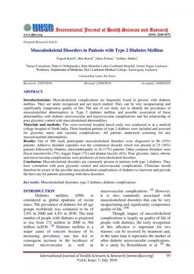
215 Introduction Musculoskeletal complications are most seen in patients with a longstanding history of DM type 1 Aim This study aimed to determine the prevalence of musculoskeletal manifestations ID: 947638

foot diabetes type musculoskeletal diabetes foot musculoskeletal type participants pain study prevalence deformities chippaux index mellitus joint patients deformity

Share:

Link:

Embed:

Download Presentation from below link

Download Pdf The PPT/PDF document "Revati J Nalawade revatijngmailcomINTROD..." is the property of its rightful owner. Permission is granted to download and print the materials on this web site for personal, non-commercial use only, and to display it on your personal computer provided you do not modify the materials and that you retain all copyright notices contained in the materials. By downloading content from our website, you accept the terms of this agreement.


Presentation Transcript

215 : Revati J Nalawade: revatijn@gmail.comINTRODUCTION Diabetes mellitus is a group of metabolic diseases characterized by hyperglycaemia resulting from defects in insulin secretion, insulin action, or both [1]. The global diabetes prevalence is estimated to be 9.3% whereas that in India ranges from 5–17%, with higher levels found in the southern Introduction: Musculoskeletal complications are most seen in patients with a longstanding history of DM type 1. Aim: This study aimed to determine the prevalence of musculoskeletal manifestations associated with DM type 1 in Belagavi district. Materials and Methods: Out of a total of 200 participants with Diabetes Mellitus type 1, 95 participants with musculoskeletal problems were assessed for range of motion, pain, and muscle strength of the most affected joint. They were also screened for HOW TO CITE THIS ARTICLE: Deepa C Metgud, Shukra D Chivate, Revati J Nalawade, Prevalence of Musculoskeletal Manifestations in Diabetes Mellitus Type 1: A Cross-Sectional Study, J Res Med Dent Sci, 2020, 8 (6): 215-220. Journal of Research in Medical and Dental Science | Vol. 8 | Issue 6 | September 2020 216 acute proximal neuropathy, proximal motor neuropathy, pyomyositis and the diffuse idiopathic skeletal hyperostosis (DISH) syndrome and finally c) disorders for which a possible association with diabetes has been proposed but not proven yet, such as The most common type of cheiroarthropathy observed is adhesive capsulitis (31%), followed by carpal tunnel syndrome (3-%), flexor tenosynovitis (28%), salaam deformity (22%), and Dupuytren’s contracture (9%) [5-7]. These manifestations may go unrecognized or simply be overlooked in daily clinical practice. Thus, clinicians should be aware of the possible musculoskeletal complications of diabetes to intervene and provide the best care for affected patients. The prevalence of musculoskeletal complications due to DM type 1 in Belagavi has not been estimated. Hence, our study aims to determine the prevalence of musculoskeletal manifestations associated with DM type 1 in MATERIAL AND METHODSA cross sectional study was conducted with an aim to estimate the prevalence of the musculoskeletal manifestation in patients with DM type I. The study was approved by the Institutional Ethical Committee before the commencement of the study. A free one-day camp was organized by the diabetic center along with pediatric physiotherapy department of a A total of 200 participants with DM type 1 in and around Belagavi were assessed by the diabetic center and a total of 95 participants with musculoskeletal problems were referred for physiotherapy evaluation. The participants were assessed for range of moti

on (ROM), pain and muscle strength of the most affected joint. The ROM of the affected joint was assessed by asking the participant to perform active movements of the joints and was graded as 1: Full and Pain Free, 2: Full and Painful, 3: Restricted and Pain Free, 4: Restricted and Painful. The participants were also interviewed for presence or absence of pain. A generalized assessment of strength was performed for the participants complaining of The participants were screened for any deformities in the upper and lower limbs. The deformities such as hallux valgus, hallux varus, pes cavus, pes planus, salaam deformity, etc. were recorded on observation. They were also interviewed for any activity limitations in functional tasks such as ambulation, bed mobility, dressing, bathing toilet activities and The objective outcomes used were chippaux index and weight distribution during gait. The chippaux index is a standard tool which was used to assess the height of the longitudinal foot arch [8]. Static footprints of the participants were collected on a piece of paper and impregnated with ink, between the subject’s foot and the paper [9]. To calculate the chippaux index, a line tangent to the internal edge of the footprint was drawn. From the point of contact of this line with the digito-planar eminence, a line was drawn that crossed the eminence through its broadest part. Then, a line parallel to the previous one was drawn, that passed through the narrowest point of the isthmus of the sole of the foot. The index was the quotient obtained on dividing the value of the isthmus line by the value of the breadth line of the forefoot and was expressed in percentage. The scores were further categorized as: Chippaux Index <21%: Tendency to cavus foot, 21% < Chippaux Index < 01%: Bormality range, Chippaux Index = 45%: Tendency to flatness and/or pronation [1-]. Following the chippaux index, the medial and lateral weight distribution during gait was also assessed. Descriptive analysis of the data was done using Ri386 3.63. Categorical variables were compared using chi-square test. P-value <-.-1 RESULTSOut of 200 diabetic patients, a total of 95(47.5%) patients (51 males, 44 female) presented with musculoskeletal problems. The mean age of these participants was 16.05 ± 6.17 years. Majority of the participants were students with a percentage of 70.53%. The mean duration of Among 95 participants, 14 (14.74%) presented with restricted and pain-free ROM, 1(1.05%) 217 showed full and painful ROM and 80 (84.21%) had full and pain-free ROM. The joints most involved were PIP and DIP 8.42%, only PIP 6.32%, MCP 1.05% and Knee joint 1.05%, suggesting that the PIP and DIP together were more s

ignificantly involved. Musculoskeletal pain was present in 85 (89.47%) participants and 94 (98.95%) showed generalized muscle weakness. Only 1.05 % of the total participants presented with difficulties in Musculoskeletal deformities were observed in 57 (60%) of the participants with diabetes mellitus type 1. The deformities seen in the lower extremity were pes cavus in 25 (43.86%) participants, hallux valgus in 19 (33.33%) participants, pes planus in 13(22.81%) participants, followed by genu valgum, genu varum and hallux valgus which was 7.02% respectively. Upper limb deformities included salaam deformity which was observed in 21 (36.84%) participants. The most frequent deformity observed was over toes, tingling in hands at night, wasting of hypothenar muscles, etc. were also commonly seen with a total prevalence of 40.35% The chippaux index was analysed for 90 participants as 1 could not be assessed due to open wound injury over foot. According to Chippaux index, 22 (23.33%) participants in the sample presented with tendency to cavus foot and 10(10.30%) with tendency to flatness or pronated foot. The remaining 56.84% participants showed chippaux index within normal ranges. The medial weight bearing of foot was observed in 17(17.89%) participants, whereas lateral weight bearing was seen in 27 Dsing chi square test with simulation it was concluded that ROM and deformity are significantly associated (p=-.-12) (Table 1). Age (p=-.244) and gender (p=-.3-1) of the participants did not have a significant association with deformities. Musculoskeletal deformities were more commonly seen in males (62.75%) than females (56.82%). The duration of diabetes did not have any significant relation with deformities (p=-.119). Cther parameters such as pain (p=-.311), muscle weakness (p=1) and activity limitation (p=1) did not show any significant association with musculoskeletal Age (in years)FemaleDura�on of DM (in years)Occupa�onStudentfarmer/ca�le rearingserviceOthers**Indicates mason, hotel worker, factory workerTable 1: Demographic details of participants with DM type 1 ROMFull and Pain FreeFull and PainfulRestricted and Pain FreeJoint involvedDIP, PIPPainPresentAbsentWeaknessPresentAbsentAc�vity Limita�onPresentAbsentTable 2: Prevalence of musculoskeletal manifestations in participants with DM type 1. 218 DeformityPresentAbsentTypeHallux ValgusHallux VarusPes PlanusPes CavusGenu ValgumGenu VarusSalaam DeformityOthers**Indicates pain in calf, pain in thigh, knee pain, etc.Table 3: Prevalence of musculoskeletal deformities in participants with DM type 1. Chippaux IndexTendency to cavus footNormality rangeTendency to �a

tness and/or prona�onWeight BearingLateralTable 4: Scores on Chippaux Index and weight bearing of foot in participants with DM type 1. FactorSub-categoryDeformityP-valuePresentAbsentAge Group*FemaleOccupa�onstudentNon-StudentDura�on of DM*ROMFull and Pain FreeRestricted and Pain Free APainPresentAbsentWeaknessPresentAbsentAc�vity Limita�onPresentAbsentTable 5: Association of different factors with deformity.The purpose of this study was to determine the prevalence of musculoskeletal manifestations in DM type 1. Studies have specified that limited joint mobility due to DM type 1, is reversible in early stages but becomes irreversible in time [11]. Hence age plays an important role. Longer diabetes duration has been considered as a risk factor for both limited joint mobility and and also shows that longer duration of diabetes is also considered as a predictor of foot abnormalities. However, the duration of diabetes (5.65 ± 4.9 years) in the current study was not associated with musculoskeletal deformities (p=-.119) [13]. A research conducted on effect of diabetes mellitus on osteoarthritis of knee states that the mechanism of diabetes mellitus on pain is unclear, however, diabetes mellitus type 1 may cause pain, in most cases by inducing a symmetrical sensory polyneuropathy in the distal part of the extremities, with prevalent loss of small fibers. Also, higher pain intensities were recorded during activities which put load on the joint as well as under resting conditions without load [10]. Pain was experienced by majority of The exact etiology of limited joint mobility is unknown, although there is evidence of soft tissue accumulation of advanced glycation end products in tissues, which may cause stiffening [15,16]. Our study showed that ROM was 219 associated with the presence of deformities in patients with DM type 1. Studies show that there is inability to fully flex or extend the fingers and sclerosis of tendon sheaths. The underlying cause is thought to be multifactorial. Increased glycosylation of collagen in the skin and periarticular tissue, decreased collagen possibly diabetic neuropathy are thought to be some of the contributing factors. The joints commonly affected in our study were the PIP and DIP joints. Flexion contractures of the fingers may develop at advanced stages. One indication of the presence of this condition is known as the “prayer sign” also known as the salaam deformity, which was observed as a common deformity in the upper extremities (32.40%) in the current study. This is seen when the patient is unable to press their palms together completely without a gap remaining between op

posed palms and fingers [0]. The prevalence of musculoskeletal deformities in our study was found to be 60%. Cur present findings considered together with those results of the literature show that foot abnormalities are more common in patients with DM type 1 [17,12]. Foot deformities are important contributory risk factors and predictive of foot ulceration, possibly by predisposing the skin to high pressure at the site of the foot deformity [18]. Hence, ankle and foot joint mobility must be evaluated and prevented to avoid further foot problems in adolescents with DM type 1 from early stages. Some studies have indicated a significant relationship between hallux valgus and pes planus feet when contrasted with the neutrally aligned foot. The feet may possibly share a similar etiology in most cases, such as a as a Studies have indicated that Chippaux Index values are lower in individuals with DM type 1, which means that they trend toward pes cavus [19,20]. Our results which show a foot structure deviation toward pes cavus are consistent with this knowledge. Individuals with DM type 1 are known to have a significantly lower ankle and foot range of motion, a lower ankle muscle strength, and a tendency to high medial longitudinal arch. Furthermore, muscle weakness progresses from distal to proximal in consequence of motor neuropathy leading to atrophy in the intrinsic muscles before the extrinsic muscles. This condition is known to cause muscle imbalance which may result in hammer toes, claw toes, and pes cavus [21]. Studies suggest that diabetic neuropathy may lead to disturbance of foot mechanics. A study examining gait abnormalities in diabetic neuropathy showed that there was there was more lateral position of the centre of pressure on the sole of the foot [22]. Majority of the patients from our study also show the presence of lateral weight bearing (28.42%) Absence of a control group and small sample size were the limitations of this study. Also, there was no follow-up done to comment on the severity of musculoskeletal manifestations occurring with CONCLUSIONThe present study concludes that, musculoskeletal manifestations occur with a greater frequency in patients with Diabetes Mellitus type 1. The prevalence of musculoskeletal deformities in patients with DM type 1 was 60%. Also, ROM and musculoskeletal deformities were significantly associated. We suggest that further studies should be conducted evaluating the association of a single parameter with musculoskeletal D. Diagnosis and classification of diabetes Saeedi P, Petersohn I, Salpea P, et al. Global and regional diabetes prevalence estimates for 2019 and projections for 2030 and 2045: Results from the international

diabetes federation diabetes Atlas. Diabetes Res Clin Tripathy JP, Thakur JS, Jeet G, et al. Prevalence and risk factors of diabetes in a large community-based study in North India: Results from a STEPS survey in Punjab, Kim RP, Edelman SV, Kim DD. Musculoskeletal complications of diabetes mellitus. Clin Diabetes 2001; Arkkila PE, Gautier JF. Musculoskeletal disorders in diabetes mellitus: An update. Best Pract Res Clin Douloumpakas I, Pyrpasopoulou A, Triantafyllou A, et al. Prevalence of musculoskeletal disorders in patients with type 2 diabetes mellitus: A pilot study. Hippokratia Larkin ME, Barnie A, Braffett BH, et al. Musculoskeletal e 1 diabetes. Diabetes Care 2014; 220 Vařeka I, Vařeková R. The height of the longitudinal foot arch assessed by Chippaux-Šmiřák index in the compensated and uncompensated foot types according Gonzalez-Martin C, Pita-Fernandez S, Seoane-Pillado T, et al. Variability between Clarke’s angle and Chippaux-Smirak Index for the diagnosis of flat feet. Colombia Búñez-Samper M, Búñez-Samper M, Biomecánica, 11.Nagesh VS, Kalra S. Type 1 diabetes: Syndromes in resource-challenged settings. JPMA. J Pakistan Med Majjad A, Errahali Y, Toufik H, et al. Musculoskeletal disorders in patients with diabetes mellitus: A cross-Mansour AA, Imran HJ. Foot abnormalities in diabetics: Prevalence and predictors in Basrah, Iraq. Pakistan  Eitner A, Pester J, Vogel F, et al. Pain sensation in human osteoarthritic knee joints is strongly enhanced by Rosenbloom AL, Silverstein JH. Connective tissue and joint disease in diabetes mellitus. Endocrinol Metab Clin Lindsay JR, Kennedy L, Atkinson AB, et al. Reduced prevalence of limited joint mobility in type 1 diabetes in a UK clinic population over a 20-year period. Diabetes Ledoux WR, Shofer B, Smith DG, et al. Relationship between foot type, foot deformity, and ulcer occurrence in the high-risk diabetic foot. J Rehab Res Develop 2005; Vermigli C, Carrington AL, Boulton A. Muscle weakness and foot deformities in diabetes. Diabetes Care 2004; Staheli LT, Chew DE, Corbett M. The longitudinal arch. A survey of eight hundred and eighty-two feet in normal children and adults. J Bone Joint Surg Am 1987; 69:426– Forriol F, Pascual J. Footprint analysis between three and seventeen years of age. Foot Ankle 1990; 11:101– Mutlu EK, Unver B, Taskiran H, et al. An investigation of the foot ankle joint mobility, muscle strength, and foot structure in adolescent with type 1 diabetes. Int J Katoulis EC, Ebdon-Parry M, Lanshammar H, et al. Gait Journal of Research in Medical and Dental Science | Vol. 8 | Issue 6 | September 2020 Deepa C Metgud, et al.J Res Med Dent Sci, 2020, 8 (6):215-220