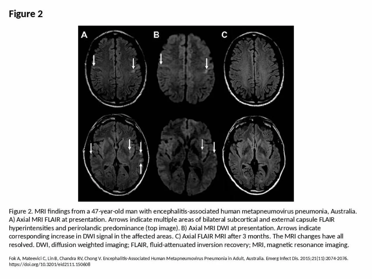
PPT-Figure 2 Figure 2. MRI findings from a 47-year-old man with encephalitis-associated human

Author : arya | Published Date : 2024-07-04

Fok A Mateevici C Lin B Chandra RV Chong V EncephalitisAssociated Human Metapneumovirus Pneumonia in Adult Australia Emerg Infect Dis 2015211120742076 httpsdoiorg103201eid2111150608

Presentation Embed Code

Download Presentation

Download Presentation The PPT/PDF document "Figure 2 Figure 2. MRI findings from a 4..." is the property of its rightful owner. Permission is granted to download and print the materials on this website for personal, non-commercial use only, and to display it on your personal computer provided you do not modify the materials and that you retain all copyright notices contained in the materials. By downloading content from our website, you accept the terms of this agreement.

Figure 2 Figure 2. MRI findings from a 47-year-old man with encephalitis-associated human: Transcript


Fok A Mateevici C Lin B Chandra RV Chong V EncephalitisAssociated Human Metapneumovirus Pneumonia in Adult Australia Emerg Infect Dis 2015211120742076 httpsdoiorg103201eid2111150608. Figure 12.1a The functions of cell division: Reproduction. Figure 12.1b The functions of cell division: Growth and development. Figure 12.1c The functions of cell division: Tissue renewal. Figure 12.2 Eukaryotic chomosomes. -1- Figure 1 -2- Figure 3 Figure 4 Figure 5loss resistance is zero, and that the radiation resistance is the textbook figure of 35 ohms. The feedpointimpedance would then be 15+0+35 = 50 ohms, and th Sixth Edition. Chapter 5. Heredity and Disease. Mary Lou Mulvihill. Mark Zelman. Paul Holdaway. Elaine Tompary. Jill Raymond. Chapter 5. . Heredity and Disease. Slide 10 Sickle Cells. Slide 24 Down Syndrome. Figure 12.1b The functions of cell division: Growth and development. Figure 12.1c The functions of cell division: Tissue renewal. Figure 12.2 Eukaryotic chomosomes. Figure 19.0 Chromatin in a developing salamander ovum. Keller C, Zumblick M, Streubel K, Eickmann M, Müller D, Kerwat M, et al. Hemorrhagic Diathesis in Borrelia recurrentis Infection Imported to Germany. Emerg Infect Dis. 2016;22(5):917-919. https://doi.org/10.3201/eid2205.151557. Figure 1.3. Figure 1.4. Radius. Ulna. Humerus. Annular . Ligament. Radial. Collateral . Ligament. Capitellum. /. Capitululm. Photo taken from: Color Atlas of Human Anatomy (1997). p. 122. Left elbow joint and proximal radio-ulnar joint, B from the lateral side.. Zajkowska J, Moniuszko A, Czupryna P, Drozdowski W, Krupa W, Guziejko K, et al. Chorea and Tick-Borne Encephalitis, Poland. Emerg Infect Dis. 2013;19(9):1544-1545. https://doi.org/10.3201/eid1909.130804. Telford SR, Armstrong PM, Katavolos P, Foppa I, Garcia AS, Wilson ML, et al. A New Tick-borne Encephalitis-like Virus Infecting New England Deer Ticks, Ixodes dammini. Emerg Infect Dis. 1997;3(2):165-170. https://doi.org/10.3201/eid0302.970209. Messenger SL, Smith JS, Orciari LA, Yager PA, Rupprecht CE. Emerging Pattern of Rabies Deaths and Increased Viral Infectivity. Emerg Infect Dis. 2003;9(2):151-154. https://doi.org/10.3201/eid0902.020083. Wangchuk S, Tamang T, Darnal J, Pelden S, Lhazeen K, Mynak M, et al. Japanese Encephalitis Virus as Cause of Acute Encephalitis, Bhutan. Emerg Infect Dis. 2020;26(9):2239-2242. https://doi.org/10.3201/eid2609.200620. Chung M, Lee S, Kim M, Lee J, Kim E, Lee J, et al. Japanese Spotted Fever, South Korea. Emerg Infect Dis. 2006;12(7):1122-1124. https://doi.org/10.3201/eid1207.051372. Basavaraju SV, Kuehnert MJ, Zaki S, Sejvar J. Encephalitis Caused by Pathogens Transmitted through Organ Transplants, United States, 2002–2013. Emerg Infect Dis. 2014;20(9):1443-1451. https://doi.org/10.3201/eid2009.131332. Saivish MV, da Costa VG, Rodrigues RL, Féres V, Montoya-Diaz E, Moreli ML. Detection of Rocio Virus SPH 34675 during Dengue Epidemics, Brazil, 2011–2013. Emerg Infect Dis. 2020;26(4):797-799. https://doi.org/10.3201/eid2604.190487. Young DS, Kramer LD, Maffei JG, Dusek RJ, Backenson P, Mores CN, et al. Molecular Epidemiology of Eastern Equine Encephalitis Virus, New York. Emerg Infect Dis. 2008;14(3):454-460. https://doi.org/10.3201/eid1403.070816.

Download Document

Here is the link to download the presentation.
"Figure 2 Figure 2. MRI findings from a 47-year-old man with encephalitis-associated human"The content belongs to its owner. You may download and print it for personal use, without modification, and keep all copyright notices. By downloading, you agree to these terms.

Related Documents