/
Subcutaneous Abdominal Fat Measurement using Ultrasonography in Indivi Subcutaneous Abdominal Fat Measurement using Ultrasonography in Indivi

Subcutaneous Abdominal Fat Measurement using Ultrasonography in Indivi - PDF document

ash
ash . @ash
Follow
342 views
Uploaded On 2022-10-12

Subcutaneous Abdominal Fat Measurement using Ultrasonography in Indivi - PPT Presentation

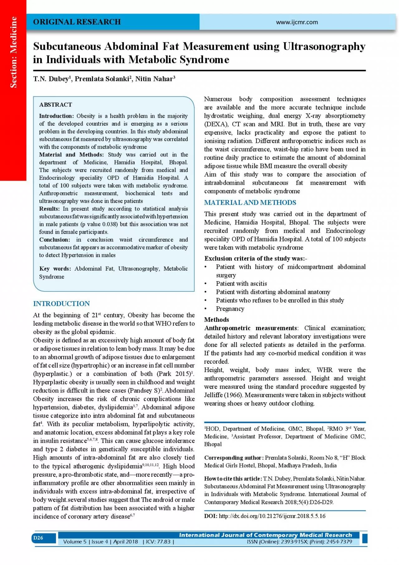
D26 Introduction Obesity is a health problem in the majority of the developed countries and is emerging as a serious problem in the developing countries In this study abdominal subcutaneous fat meas ID: 959276

fat x00660069 study obesity x00660069 fat obesity study waist subcutaneous abdominal bmi metabolic signi disease patients male cardiovascular measured

Share:

Link:

Embed:

Download Presentation from below link

Download Pdf The PPT/PDF document "Subcutaneous Abdominal Fat Measurement u..." is the property of its rightful owner. Permission is granted to download and print the materials on this web site for personal, non-commercial use only, and to display it on your personal computer provided you do not modify the materials and that you retain all copyright notices contained in the materials. By downloading content from our website, you accept the terms of this agreement.


Presentation Transcript

D26 Subcutaneous Abdominal Fat Measurement using Ultrasonography in Individuals with Metabolic SyndromeT.N. Dubey, Premlata SolankiL RE Introduction: Obesity is a health problem in the majority of the developed countries and is emerging as a serious problem in the developing countries. In this study abdominal subcutaneous fat measured by ultrasonography was correlated At the beginning of 21 century, Obesity has become the leading metabolic disease in the world so that WHO refers to Obesity is de�ned as an excessively high amount of body fat or adipose tissues in relation to lean body mass. It may be due to an abnormal growth of adipose tissues due to enlargement of fat cell size (hypertrophic) or an increase in fat cell number (hyperplastic.) or a combination of both (Park 2015)Hyperplastic obesity is usually seen in childhood and weight HOD, Department of Medicine, GMC, Bhopal, www.ijcmr.com International Journal of Contemporary Medical Research Volume 5 | Issue 4 | April 2018 | ICV: 77.83 |ISSN (Online): 2393-915X; (Print): 2454-7379 Section: Medicine D27 was measured to the nearest centimetre using an anthropometric inelastic measuring tap with the subject Weight of the subjects was measured by weighing machine. Waist and hip measurements were taken using an inelastic The amount of subcutaneous and abdominal adipose tissue was measured anthropometrically and ultrasonographically. The subjects’ height and weight were measured while they was calculated as weight (kg) Weight (Kg.)Normal range of BMI is considered in between 18.5 to 22.9. BMI 23 or more than 23 is considered as at risk(WHO Waist circumference was measured halfway between the lower rib and the iliac crest, and hip circumference was measured at the level of the greater trochanter (10): In males, we used waist circumference cut of�fs at 90cm while in females, the cut of�fs used were at 80cm for Indian subjects.(International diabetes federation global consensus de�nition)In making the ultrasound measurementsOf abdominal subcutaneous fat, Measurement of subcutaneous fat was performed with the patient in dorsal decubitus position and the convex 3-4 MHz transducer cross sectionally placed on midline,1cm above the umbilicus, during the expiratory phase without pressure on the abdomen in order not to distort measurement subcutaneous fat corresponds to distance in cm between the skin ant anterior surface of the linea alba which is the tendinous raphe that Waist hip ratio was measured as:-Waist circumferenceNormal waist hip ratio for Indian male is female it is (gy Task Force Consensus Group.LANCET 2005;366:1059-69)Biochemical test(lipid pro�le) was performed in departm

ent Patients with Serum Triglyceridedepartment level 150mg/dl and males with HDL with HDL in STATISTIData were entered in excel worksheet and analysed using stastical package for social science stastcal softwere for window (SSPE version 17) difference was considered The present study was undertaken to correlate intraabdominal iiiLowNormaleeeHighNormalPresentPresentTable-1: PresentPresentCorrelation OF BMI with HDL HTN and DM2 anthropometry with other components of metabolic syndrome 100 patients with either symptomatic disease (CVA, CAD, peripheral arterial disease or a marked cardiovascular disease risk factors like HTN, dyslipidemia, or DM) were In our study BMI was not signi�cantly related to any of component of metabolic syndrome P value was statistically insigni�cant other studies also declare the superceding of Two indicator were studied purposely to measure abdominal obesity –WHR and WC.In our study male participants showed statistically signi�cant correlation between waist circumference and hypertention but this association was not shown by the female participants (p value 0.045) waist hip ratio could not be associated Several studies are also in favour of waist circumference as a stronger predictor for metabolic risk factors and cardiovascular disease as compare to WHR.According to statistical analysis subcutaneous fat was signi�cantly associated with hypertension in male patients But this correlation was not found to be signi�cant in females The present study was undertaken to compare the association of ultrasonography and waist circumference measurement of abdominal subcutaneous fat with other component of metabolic syndrome among patients attending hamidia In this study patients visiting hamidia hospital were taken with either symptomatic cardiovascular disease (cerebral ischeamia, coronary artery disease, peripheral artery disease, or abdominal aortic aneurysm) or a marked cardiovascular In this study study population was strati�ed in to 10 years of A total of 1 (1%) subjects were in the age group of of age. Age group 31-40 consist of 18 (18%) subjects.Age group 41-50 constitute highest no of population i.e.34 5 female and 9 male. 0 y;êrs;&#x 000;Age group 70 years consists of 4 In our study, prevalence of overweight (BMI23-29.9) was 51. 37 patients were put in obese class 1 (BMI 25-29.9) and 7 patients were found to be class 2 obese (BMI30.0-34.9) and 4 patients were included in the group of class 3 obese patient (BMI35.0 -39.9). (TABLE NO -2)In this study the BMI was ranged between 22.08 - 43.2 in females and ranged between 23.43 - 40.90 in males. Mean BMI of the study popu

lation was 30.07±4.04, females were comparatively more obese than males. De�ning obesity in PresentPresentCorrelation of Whr with HDL HTN and DM2 Waist circumferencePresentPresent101-110�110101-110�1100.11Correlation of waist circumference with HDL, TG, HTN and DM Dubey, et al.Subcutaneous Abdominal Fat Measurement using Ultrasonography Section: Medicine International Journal of Contemporary Medical Research Volume 5 | Issue 4 | April 2018 | ICV: 77.83 |ISSN (Online): 2393-915X; (Print): 2454-7379 relation to BMI (cut of�f value 25gm/mIn our study BMI was not signi�cantly related to any of Visscher TL et al. total 6296 men and women were studied they found that BMI is a poor predictor of found that BMI is a poor predictor of cardiovascular risks and mortality than WHR and WC.1.01 ±0.10 males were comparatively having more WHR In our study abdominal obesity according to waist hip ratio, was found in 98 individuals. We didn’t �nd any signi�cant correlation between WHR and component of metabolic We found a signi�cant correlation between waist circumference and hypertention in male patient (p value 0.045) (table no 4) but this was not found to be signi�cant in In our study Ultrasound Intraabdominal fat measurements were performed in 100 participants and mean value of for female was 1.67±0.67 and mean cutaneous fat for male was 1.56±0.62. according to statistical analysis subcutaneous fat was signi�cantly associated with hypertension in male But this correlation was not found to be signi�cant in females patients (p value 0.38). (Table NO -1).Similar study was conducted in pril 2003 by Ronald P stalk et al they analyse 600 participants. they found that subcutaneous fat measured by ultrasound was not signi�cantly related to the presence of metabolic syndrome. Table-1,2,3,4 provides the association between three anthropometric indices and subcutaneous fat with DM/HTN/Obesity-associated arterial hypertension is characterized by activation of the sympathetic nervous system, activation of the renin-angiotensin system, and sodium retention, among More recent evidence indicate that upper body obesity imparts a greater risk for cardiovascular disease than lower Praxis et al French study conducted in1996 that obesity of the male (Android obesity) is clearly a cardiovascular risk factor, more so than gynecoid obesityAnother hypothesis of androgen and oestrogen receptors suggest that adipocytes have speci�c receptors for androgens, unlike most hormones testosterone induces an increase in no of androgen receptors after exposure to fat cells, thereby affecting lipid

mobilization.have the same characterstics as that found in male adipose tissue. however, estrogen treatment down regulate the density of this receptor, which might be a mechanism whereby oestrogen protect adipose tissue from androgen effect. strogen by itself seems to protect postmenopausal women receiving replacement therapy.In conclusion waist circumference and subcutaneous fat appears as accommodative marker of obesity to detect Hypertension in males. However as this study is hospital based, a community based cohort study with representative sample is required to identify a sensitive and contextual marker in Indian sub-continent which will enable us to intervene in pre pathogenesis phase and to detect at earliest the cardiovascular morbidity.Park textbook of preventive and social medicine. K. Park, Pendesey S. Practical management of diabetes. 251-270; Anjana, M.; Sandeep, S.; Deepa, R.; Vimaleswaran, K. S.; Farooq, S.; Mohan, V. Visceral and Central Abdominal Fat and Anthropometry in Relation to Diabetes in Asian Poehlman, Eric T. bdominal Obesity: The Metabolic Multi-risk Factor.Coronary Heart Disease. Exp. 1998;9: Després, Jean-Pierre; Lemieux, Isabelle. Abdominal Obesity and Metabolic Syndrome. Nature 2006;444:881–Expert Panel On Detection, Evaluation. xecutive Summary of The Third Report of The National Cholesterol Education Program (NCEP) Expert Panel on Detection, Evaluation, And Treatment of High Blood Cholesterol In Adults (Adult Treatment Panel III). JAMA: the Journal of the American Medical Association 2001;285: 2486–97.Grundy SM, Brewer HB, Cleeman JI, Smith SC, Lenfant D, for the Conference Participants. De�nition of metabolic syndrome: report of the National, Heart, Lung, and Blood Institute/American Heart Association conference on scienti�c issues related to de�nition. Visscher Tl et al. underreporting of bmi in adults and its effect on obesity prevalence period estimation Schnieder HJ et al. The predictive value of different measures of obesity for incident cardiovascular events and mortality. J Clin Endocrinol Metab 2010;95:1777-85. Ronald P Stolk et al. Ultrasound measurements of intraabdominal fat estimate the metabolic syndrome better than do measurement of waist cirference. Am J 11.Who Consultation of Obesity. Preventing and managing of global epidemic. Geneva; World health organigation Janjic D. Android type obesity and gynoid type obesity. Source of Support: Con�ict of Interest: Dubey, et al.Subcutaneous Abdominal Fat Measurement using Ultrasonography Section: Medicine International Journal of Contemporary Medical Research ISSN (Online): 2393-915X; (Print): 2454-7379 | ICV: 77.83 |Volume 5 | Issue 4 | April 2